Diseases of inflammatory etiology are among those that occur in pets not so rarely. One of them is eosinophilic granuloma in cats. It is accompanied by an inflammatory process on the pet’s skin and oral mucosa, which is most often caused by an allergic reaction to the bites of parasites such as fleas or mosquitoes, as well as hypersensitivity to certain components of the daily diet.
Eosinophilic granuloma complex is so named because of the immune cells that are classified in veterinary medicine as eosinophils (a type of white blood cell). According to experts, they are the initiators of the disease. The article will discuss the main causes and symptoms of the disease, as well as offer options for its effective treatment and prevention.
Why does EEG occur?
The cause of EEG may be an allergy to flea bites or other ectoparasites, a reaction to food components, or environmental allergens, such as pollen or house dust.
Research has shown that in at least some (rather rare) cases, the tendency to develop eosinophilic skin diseases is genetically inherited. It has been suggested that genetic predisposition and environmental influences may play a joint role in the manifestation of eosinophilic skin diseases. The hypothesis that these pathologies are rarely caused by only one of the factors explains why EEG can occur during treatment or persist for several months or even years.
Diagnostics
Most experienced veterinarians will be able to determine the form of the pathology only by the appearance of the cat, which will help prescribe the correct treatment. However, it is better to make a diagnosis only after a biopsy and cytological sampling. This is done in order to exclude the oncological nature of the tumor.
A prerequisite for differential diagnosis will be:
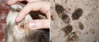
- scraping from the site of the cat's skin lesion;
- exclusion of bacterial infection using cytological examination;
- therapy against ticks and fleas;
- a special diet that will help determine whether the cat’s diet contains the allergen causing the rash.
Only after carrying out all the above manipulations can we say with confidence that the pet will be treated correctly for the type of illness that causes such a reaction in the body.
Types of CEG
Three types of skin lesions have been described in eosinophilic granuloma complex:
- An eosinophilic plaque is a hairless, flat, raised lesion that is often red, moist, and shiny. Plaques are most often found in the groin and armpits or on the outer thigh. These lesions are often itchy and cats may lick them constantly.
- Eosinophilic granuloma is a hairless, raised, often whitish-yellowish lesion on the skin or mouth, often with ulcers on the surface. Most often, granulomas are found on the back of the thighs (you can find the name “ linear granuloma ”), they can also occur in the oral cavity and on the tongue. Less commonly, other parts of the body (chin, paw pads, etc.) may be affected.
- An indolent ulcer is a well-defined defect (erosion or ulcer) on one or both upper lips. First, the lip swells, then an ulcer forms. The ulcer is usually painless and does not bleed.
Feline eosinophilic granuloma complex
Svetlana Belova, Estonian University of Natural Sciences, www.vetderm.eu Photos of the author are used in the article.
Introduction
The eosinophilic granuloma complex (EGC) includes three components, namely: an ulcer on the upper lip, also called an indolent ulcer, an eosinophilic plaque and an eosinophilic granuloma. These three components and the entire complex as a whole are not a specific diagnosis, they are just various clinical manifestations, a kind of inflammatory reaction of the skin to a primary disease, which, as a rule, is of allergic etiology.
The components of a CEG can occur both together (as a complex) or separately, are not always characterized by an eosinophilic infiltrate (for example, an indolent ulcer), and do not always form a granuloma, so the name itself (CEG) has been criticized more than once and it has even been proposed to rename the CEG to eosinophilic dermatoses of cats, but the new name has not yet found widespread use.
EEGs are dermatoses commonly found primarily in young adult (average age of onset: 3.5 years) cats. There is no proven breed or gender predisposition. Although, judging by the results of one study, there may be some genetic basis for the occurrence of eosinophilic granulomas.
Etiology
In the vast majority of cases, signs of EEG are caused by primary hypersensitivity reactions - allergies to insect bites (fleas and mosquitoes, mainly), food allergies or atopy - hypersensitivity to airborne allergens.
Clinical picture
An indolent ulcer (IU) is a clearly defined defect (erosion or ulcer, depending on the stage of the disease) with raised edges on one or both upper lips (photo 1, 2). The defect affects not only the skin, but also the mucous membrane of the oral cavity. There is no pain or itching, and the ulcer does not bleed. The surface of the IV may appear yellowish-brownish, with whitish inclusions. The lip itself, due to the inflammatory infiltrate accumulated in the thickness of the skin, is enlarged and swollen. Most likely, although there are different opinions on this matter, the ulcer is caused by increased licking of other parts of the body due to itching.
| Photo 1. Bilateral indolent ulcer at an early stage of development | Photo 2. Unilateral indolent ulcer, chronic lesion |
Eosinophilic plaque (EP) is a clearly circumscribed lesion raised above the skin level with a moist, shiny, erosive surface (photos 3, 4, 5). It can be located anywhere, but the classic localization of EB is the abdomen, groin, inner and back of the thighs. EB is characterized by severe itching.
Eosinophilic granuloma (EG) can be located in the oral cavity (whitish papules or nodules, sometimes with erosions, usually on the tongue and palate, photos 6, 7), on the chin (swelling, sometimes with a shiny, hairless surface, the so-called “fatty” chin”, photo 8), on the paws (hard itchy formation in the interdigital spaces or in the thickness of the paw pads, photo 9) or on the back surfaces of the thighs (the so-called linear or linear granuloma, a lesion in the form of a dense, rope-like intradermal thickening, photo 10 ). Linear granuloma can sometimes be found on the sides or abdomen (photo 11).
| Photo 3. Multiple eosinophilic plaques, classic localization | Photo 4. Wet erosive surface of eosinophilic plaque |
| Photo 5. Severe case of allergic dermatitis - eosinophilic plaques distributed over the entire ventral surface of the body | Photo 6. Multiple EGs on the lower surface of the tongue |
| Photo 7. Severe case of EEG - eosinophilic granulomas on the palate and indolent ulcers on both lips. Photo courtesy of Candace Sousa | Photo 8. Eosinophilic granuloma on the lower lip |
| Photo 9. Enlarged palmar paw pad due to infiltration of EG in its thickness | Photo 10. Linear EG with an erosive surface on the caudal part of the thigh |
| Photo 11. Two linear EGs located parallel on the side of a cat with flea allergic dermatitis |
Diagnosis
Clinical signs are quite characteristic, and in most cases cytological examination is sufficient to confirm the diagnosis of EEG. A smear from the surface or a fine-needle biopsy from the thickness of the EB or EG, as a rule, contains a large number (50% or more) of eosinophils, and in addition - neutrophils and, possibly, bacteria. It must be borne in mind that cytological material taken from the surface of the ulcer or chronic EB is unlikely to be rich in eosinophils. Due to secondary bacterial infection, the preparation will have a predominant majority of neutrophils and macrophages, as well as microbes (usually staphylococci).
In some cases, histological examination of the lesion may be necessary, for example to exclude a tumor - in particular squamous cell carcinoma - in the case of ulcerations on the lips, or to exclude herpes viral dermatitis in the case of erosive plaques, especially those located on the muzzle.
Eosinophilia in the blood can often be observed in cats with EB and EG. However, this is not a reason to believe that allergies in cats should be diagnosed by a blood test, since a cat with allergies may not have eosinophilia and, conversely, the latter is not a pathognomic sign of skin allergies and can also occur in other diseases (for example, asthma or endoparasitosis).
Treatment
Treatment consists primarily of finding and controlling the primary cause of EEG (see above). Symptomatic treatment can be aimed at suppressing itching and the inflammatory response - this is usually either glucocorticosteroids (for example, prednisolone or methylprednisolone 1-2 mg/kg per day for 5-14 days, then gradually reducing the dose to the minimum effective) or cyclosporine (5 –10 mg/kg per day). If a secondary bacterial infection is present, and chronic lesions are usually deeply infected, systemic antibiotic therapy (eg, amoxicillin with clavulanic acid 15–25 mg/kg twice daily) may be necessary.
Bibliography
1. Power HT, Ihrke PJ Selected feline eosinophilic skin diseases. Vet Clin North Am Small Anim Pract. July 1995; 25(4):833–850.
2. Muller and Kirk's Small Animal Dermatology, 6th Ed. Scott DW, Miller WH, and Griffin CEWB Saunders, 2001.
3. Fondati A., Fondevila D., Ferrer L. Histopathological study of feline eosinophilic dermatoses. Vet Dermatol. 2001 Dec; 12 (6): 333–338.
4. Bardagi M., Fondati A., Fondevila D., Ferrer L. Ultrastructural study of cutaneous lesions in feline eosinophilic granuloma complex. Vet Dermatol. 2003 Dec; 14(6):297–303.
5. Vercelli A., Raviri G., Cornegliani L. The use of oral cyclosporin to treat feline dermatoses: a retrospective analysis of 23 cases. Vet Dermatol. 2006 Jun; 17 (3): 201–206.
6. Wildermuth BE, Griffin CE, Rosenkrantz WS Response of feline eosinophilic plaques and lip ulcers to amoxicillin trihydrate-clavulanate potassium therapy: a randomized, double-blind placebo-controlled prospective study. Vet Dermatol. 2012 Apr; 23 (2): 110–118.
7. Buckley L., Nuttall T. Feline eosinophilic granuloma complex(ities): some clinical clarification. J Feline Med Surg.
SVM No. 1/2013
Rate material
Like Like Congratulations Sympathy Outrageous Funny Thoughtful No words
4
If your cat is diagnosed with EEG
Skin lesions caused by eosinophilic granuloma complex may wax and wane. If the root cause is identified and brought under control, then the lesions may disappear and not recur.
For example, it turns out that your pet is allergic to flea bites, so he is intensively treated with flea sprays or drops, and the symptoms go away. And if a cat is allergic to food containing beef protein, all types of products containing this protein are excluded from the diet.
In case of confirmed food intolerance to any food components, the veterinarian may prescribe a medicinal line of food.
For some cats, the problem may be seasonal. Seasonality is usually associated with reactions to flea, mosquito, and other insect bites or reactions to environmental allergens (such as pollen). You may need to consult a veterinary dermatologist to determine the cause of EEG.
Causes of pathology
It should be said right away that science has not established the absolutely exact factors that cause the development of eosinophilic granuloma in cats. However, statistical data suggest that there are reasons that provoke the development of this disease to a greater extent. Here they are:
- Sex and breed predisposition. Cats, due to the fact that hormonal disruptions occur more often in their bodies, are more susceptible to this disease than cats. Norwegian forest cats are particularly vulnerable to this disease, but it often occurs in other breed lines.
- Bad genetics of the pet.
- Nonspecific and food allergies in a pet.
- Dermatitis caused by tick and flea bites.
- Cat hypersensitivity.
Despite the abundance of possible causes, the owner needs to know that granuloma, in most cases, is a specific manifestation of a general or local allergic reaction to an internal or external irritant.
Drugs for the treatment of dermatitis in cats caused by EEG
Treatment is aimed primarily at finding out the cause and bringing it under control. Symptomatically used drugs to reduce itching are steroids (for example, Prednisolone) or Cyclosporine. For deep ulcers and secondary infection, antibiotics may be required.
The situation is strictly individual in each case, therefore, if the symptoms listed above appear, you should absolutely not self-medicate. It is imperative to consult a veterinarian to make a correct diagnosis. After all, even what outwardly looks like an EEG can turn out to be a more serious disease, even a malignant tumor.
Pet treatment
To accurately determine the diagnosis of granuloma in kittens, the veterinarian will examine the pet and, if necessary, conduct a microscopic examination (cytology). Based on the cytology results, the specialist will rule out other diseases with similar symptoms and perform a wet test to identify fleas and ticks. It often happens that it is impossible to determine the origin of the disease. Then complex treatment is applied. Local therapy for granuloma in cats is useless. Antiparasitic agents must be administered, regardless of whether the kitten has them or not. Only after this is it allowed to carry out anti-inflammatory therapy. Treatment is carried out mainly with the following steroid hormonal agents:
— Prednisolone; — Dexamethasone; - Triamcinolone.
Treatment is carried out for 21 days with tablets. If giving pills to a kitten is problematic, then you can replace them with injections. In this case, injections are given twice with a break of a week. Additionally, to reduce allergic symptoms, one of the following drugs is prescribed:
- Diphenhydramine; — Chlorambucil; - Cyclosporine.
In severe cases of the disease, a course of antibiotics is prescribed. After 1-1.5 weeks, the condition of a kitten with granuloma will improve significantly, but treatment cannot be interrupted. For older cats, therapy is increased if necessary to eight weeks.
Pyogranulomatous inflammation. Sterile granuloma syndrome
Sterile granuloma/pyogranuloma syndrome is a skin disease of a presumably immune-mediated nature that manifests itself in the formation of granulomatous or pyogranulomatous nodules and plaques in the skin in the absence of infectious pathogens.
The exact cause and pathogenesis of the disease have not been determined; it is presumably an immune dysfunction or an aberrant response to an antigen of an unidentified pathogen, or the initiation by the antigen of a given pathogen of a special form of immune response.
Clinical signs.
In dogs, sterile granuloma/pyogranuloma syndrome is quite rare and can occur in dogs of any age, breed and gender, with breeds such as collie, dachshund, Doberman, English bulldog, Great Dane, boxer and golden retriever likely to be predisposed. Typical locations for lesions are the head (especially the bridge of the nose, lips and skin around the eyes), ears and fingers, but lesions are also likely to spread throughout the body. The disease is characterized by the formation of multiple hard papules, plaques and nodules, which are often hard and painless, but in some cases their surface becomes ulcerated and becomes secondarily infected.
In cats, unlike dogs, the disease is extremely rare; skin lesions are characterized by the formation of multiple hard papules, plaques and nodules on the head, lips, and ears. In rare cases, skin lesions are observed on the fingers; the skin of the torso is extremely rarely affected.
Cytological examination reveals pyogranulomatous or granulomatous inflammation in the absence of infectious pathogens. Dermatohistopathological examination reveals diffuse or nodular granulomatous or pyogranulomatous dermatitis, also in the absence of microorganisms.
Diagnosis The final diagnosis is established on the basis of characteristic clinical signs, histopathological and cultural studies. The main group of differential diagnoses are granulomatous and pyogranulomatous lesions (bacterial, fungal, foreign body reaction) and neoplasms. Recently, PCR diagnostics has played an increasingly important role in diagnosis, and in areas where leishmaniasis is endemic, this type of research plays a significant role in excluding leishmaniasis as the cause of the formation of granulomas.
Treatment
At the beginning of therapy, it is optimal to carry out a trial treatment with drugs of the tetracycline group, and only after an unsuccessful attempt of this method can one move on to more powerful immunosuppressive drugs. In the absence of effect from tetracycline group drugs, either surgical excision of single lesions (if possible) or therapy with corticosteroids and non-steroidal immunosuppressive drugs is performed.
The prognosis for this disease is generally favorable, but lifelong maintenance therapy may be required.
Share
Granuloma treatment methods
For each type of disease, special therapy is indicated, so you cannot treat granuloma on your own. For the treatment of inflammatory nodules of infectious origin, appropriate medications are used, the action of which is aimed at the causative agent of the infectious disease. Antibiotics, immunomodulators, and vitamins can be used as medications.
Sometimes they resort to radiation therapy, cryotherapy and chemotherapy. Surgical removal of granulomas is also possible. Laser therapy is considered one of the most effective methods.
It is better to start treatment of granuloma at the earliest stages of the disease. Sometimes the course of treatment can last up to several years. It is very important to adhere to a diet; the patient’s nutrition should be complete and balanced.
Types of granuloma
Depending on the cause of occurrence, granulomas are divided into:
- infectious (associated with bacteria, viruses);
- non-infectious (formed around foreign bodies);
- of unknown etiology (with Crohn's disease, sarcoidosis, etc.).
In addition, the following types of granulomas are distinguished::
- pyogenic – chronic formation caused by staphylococcal infection; localized on the face, legs, fingers and arms, often resulting from skin injuries;
- ring-shaped - a collection of small nodules arranged in the form of a ring; in most cases it is localized on the neck, hands, buttocks, knees and feet;
- tuberculosis - is a zone of necrosis, which is surrounded by epithelial cells and leukocytes; can be localized on the skin, lymph nodes, lungs, respiratory tract;
- purulent - most often located on the hands or face, mainly found in children; often these granulomas occur after wounds, but the exact cause of their appearance is unknown;
- scleroma - localized mainly on the mucous membrane of the upper respiratory tract, can lead to their stenosis (narrowing) or asphyxia;
- migrating subcutaneous granuloma - its location can vary, in most cases it is localized on the lower jaw and is a complication of osteomyelitis or chronic periodontitis;
- venereal - occurs against the background of sexually transmitted infections, for example, syphilis, chlamydia, is localized in the groin and genital area, occurs mainly in men;
- ligature (postoperative) – develops as a reaction to suture material after surgery.
Symptoms of granuloma
Since most granulomas are not an independent disease, symptoms can be varied and depend on the background of the pathology they appeared against. Sometimes the first signs may go unnoticed.
Inflamed nodules first look like bright red spots. Next, a pink papule appears. In most cases, granuloma is not accompanied by discomfort. Often it can disappear and reappear on its own.
Nodules can appear on the skin, muscles, on the walls of blood vessels, bones, teeth, mucous membranes, and internal organs. Quite common is umbilical granuloma, which appears at the site of umbilical cord removal. It can also occur in adults due to wearing piercings.
Symptoms will vary depending on the location of the lesion.:
- if the skin is affected, itching, swelling, redness may occur, and peeling of the skin may also occur;
- if the liver is damaged, there may be no symptoms, sometimes dull pain occurs in the right hypochondrium, and in rare cases jaundice may appear;
- if the lungs are damaged, a cough and shortness of breath appear; with tuberculosis, sputum may be produced;
- when teeth are damaged, toothache, bleeding gums, and discharge of pus occur;
- in case of brain damage, symptoms depend on the location of the granulomas: paralysis, deterioration of vision, hearing, and smell may be observed.
Causes of granuloma
The reasons for the occurrence of this type of formation can be different, the main ones are:
- damage to the body by infectious diseases (syphilis, typhoid fever, typhus, tuberculosis);
- collagen diseases (rheumatism, scleroderma);
- drug effects;
- diseases caused by dust (silicosis, talcosis, asbestosis);
- dental diseases.
The mechanism of granuloma formation can be divided into 4 stages:
- A collection of young cells prone to phagocytosis (a process by which cells take up and digest particulate matter).
- Active proliferation of formed phagocytes.
- Transformation of phagocytes into epithelial cells.
- A collection of epithelial tissue that forms a granuloma.
Treatment of eosinophilic granuloma in cats
In most cases, eosinophilic granuloma is successfully treated. But, in case of extensive damage, the animal experiences discomfort and pain; sometimes hospital treatment is recommended.
The first thing you need to do is start a diet that excludes allergic reactions.
In addition, you should start using anti-inflammatory hormonal drugs.
It is also possible to take antibiotics to combat concomitant infections.
And also local treatment of the affected areas with special means.
The most important and important thing is to seek help in a timely manner and unquestioningly use the means prescribed by the veterinarian.
Prevention
You can prevent a cat disease such as eosinophilic granuloma if you carefully monitor the health of your pet. First of all, you need to protect him from possible allergic reactions, as a form of hypersensitivity to flea or tick bites, or food components. Doing this is not as difficult as it seems at first glance. It is enough to buy your cat high-quality, hypoallergenic food and regularly check the skin and treat it with acaricidal agents.
Such preventative measures, along with hygiene and vaccinations, will help the cat stay healthy, and owners will not have to worry about the fact that their beautiful pet will be disfigured by an ugly granuloma.
Diagnosis of eosinophilic granuloma in cats
To make a diagnosis of eosinophilic granuloma, it is necessary to conduct a diagnostic examination of the animal to exclude other diseases with a similar clinical picture. The veterinarian has at his disposal all the necessary equipment and highly qualified specialists to accurately make a correct and timely diagnosis.
The veterinarian must conduct a comprehensive examination of the cat, at the same time, much depends on the owner of the animal, because he needs to be given full information about the first symptoms, about the possible causes of the disease (flea and tick bites or allergies).
A comprehensive method for diagnosing eosinophilic granuloma includes:
- general and biochemical blood test (to identify an increased number of eosinophils);
- skin biopsy (to exclude tumor formations);
- microscopy of skin scrapings (to confirm or exclude the presence of skin parasites).