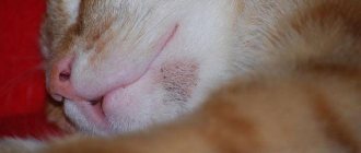
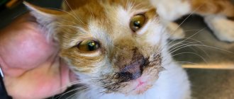
As in humans, acne in cats is a fairly common skin disease characterized by an inflammatory process in the sebaceous glands. Treatment of the disease should not be ignored under any circumstances, since acne and blackheads that appear, often on the pet’s chin, cause him a lot of discomfort.
How can you treat acne in cats?
In the fight against this disease in four-legged furry patients, areas of inflammation are sometimes treated with a two-phase liquid makeup remover. By carefully soaking a cotton swab in this product, you need to remove dirt and excess sebum - this is an excellent disinfection. This method is used only if there is no inflammation or pustules. Also, in the absence of wounds and ulcers, the use of folk remedies is allowed - you can treat acne with a composition of two furatsilin tablets mixed with pumpkin juice or boiled chamomile. In this case, lotions made from medicinal herbs – yarrow or celandine – can be effective.
In cases where acne is caused by allergies, switching to a different food or replacing plastic utensils with other materials - earthenware or metal - can help.
Varieties
Unnoticed blackness in the beard area of a pet in time can lead to the formation of purulent wounds, in place of which a crust will subsequently form. Fungus, bacteria and other pathogenic microorganisms grow in the wound, causing a purulent-inflammatory process. The table shows the main types of sores in cats in the chin area:
| Type of ulcers | Peculiarities |
| Surface | Are the result of scratches or shallow bites |
| The skin becomes reddish and a white dot appears on it | |
| Mostly the abscess opens on its own and does not require any treatment. | |
| Deep | Manifest against the background of bites with damage to the deep layers of the epidermis |
| The damaged surface of the skin is very painful, but in appearance it is not very different from healthy | |
| When palpating, a compacted area is noticed | |
| Spicy or hot | The inflammatory response develops quickly |
| There is a red wound, in place of which a crust appears | |
| Cold | Accompanied by a deep abscess, which has a chronic course |
| Such ulcers are permanent and may not go away for several years. | |
| Often recur | |
| Benign | Purulent discharge contains many living leukocytes |
| The cat's immune system rejects the affected areas, resulting in thick, white pus that does not have a strong odor appear on the chin. | |
| Malignant | White blood cells in the exudate die off or are very weak, while the cancer cells are strong |
| If purulent spots are not eliminated in a timely manner, phlegmon develops. |
Cat acne on the chin
Acne on the cat's chin is one of the most common skin diseases in cats of any gender and age. Symptoms range from almost invisible comedones to extremely inflamed ones. Depending on the course of the disease, cats behave differently. Some people don't react at all to the appearance of pimples, while others experience itchy pain and get nervous.
Where acne appears on the chin, hair loss and redness of the skin can be observed. At the first stage, you may notice a slight “dirtyness” - this effect is created by black dots between the hair follicles. Often everything is limited to this symptom, but sometimes it can turn into swollen red lumps that rupture and bleed.
Causes and mechanism of disease development
The reason for the failure of the keratinization process of epidermal cells in cats has not been established. Therefore, experts identify several factors that contribute to this phenomenon and lead to the appearance of black spots on the fur of cats. These include:
- increased secretion from the sebaceous glands of the epidermis;
- sticking of food debris to the beard or skin on the cat’s chin;
- decreased immunity after severe infectious diseases, as well as during the rehabilitation period after injuries or operations;
- dysfunction of the endocrine glands;
- intoxication phenomena caused by diseases of the gastrointestinal tract, liver and pancreas;
- allergic reactions;
- skin pathologies;
- dermatophytoses;
- demodicosis;
- improper care of the animal's skin and fur;
- some infectious and parasitic diseases;
- severe or prolonged stress.
Important! Owners must carefully monitor the cleanliness and integrity of the dishes from which the animal eats and drinks. Pathogenic microorganisms accumulate in chips, scratches and cracks, then enter the pet’s skin and can easily provoke inflammation and cause sores on the chin of cats.
The mechanism of comedones formation in cats occurs in 5 stages:
- Increased secretion from the sebaceous glands (sebum).
- Blockage of the ducts of the sebaceous glands with a mixture of sebum and dead epithelial cells.
- Formation of open cysts (comedones) at the mouth of the hair follicle.
- Oxidation of the contents of comedones in the presence of oxygen. Visually, this is marked by the appearance of black dots on the cat’s skin.
- Opening of comedones.
If comedones open into the external environment during injury by an animal, infection occurs and purulent inflammation develops. But sometimes cysts greatly increase in size, become inflamed and open into the dermal cavity. In this case, surrounding tissues are involved in the process. The cat's immune system tries to prevent the spread of pathogenic microflora and releases a huge number of white blood cells. When they die, they form purulent exudate, which accumulates in the internal cavity and then tends to come out. Clinically, this is manifested by the formation of abscesses, severe itching and even pain. Scratching the abscess leads to secondary infection, the formation of non-healing ulcers, which, when dry, form scabs on the cat’s chin.
Important! Attempts by cat owners to open comedones or abscesses on their own without following antiseptic rules often lead to re-infection and the development of complications.
Effective treatment: ointments for acne in cats
Obviously, for recipes to combat acne in our pets, we need to turn to professionals. Local remedies are usually prescribed, which can be quite effective if used regularly - once every one to three days, or as needed. This is primarily mupirocin in the medicinal form of an ointment or cream. A gel with 2.5% benzoyl peroxide is also used - keep in mind that this product may cause irritation in some cats. Another drug for cat acne is a gel with a 075% metronidazole content, as well as a lotion or cream with a 0.01% -0.025% tretinoin content. Local preparations containing tetracycline, clindamycin, or erythromycin have also proven effective.
Necessary treatment
If you notice a black chin on a cat or an area of the skin is covered with ulcers, then you should immediately show your pet to a veterinarian and take therapeutic measures. The beard is treated with the medications presented in the table:
| Medication group | Name |
| Antiseptic drugs | "Chlorhexidine" |
| "Miramistin" | |
| Hydrogen peroxide | |
| Shampoos for cat hygiene | "Bifar" |
| "Hartz" | |
| "Doctor" | |
| "Lactoderm" | |
| Medicines against ulcers in the form of ointments and gels | "Flucinar" |
| "Synthomycin liniment" | |
| Sulfuric ointment |
The affected area is washed with a decoction.
If crusts or spots have formed in the fur, it is possible to get rid of them by using iodine. Medicines based on natural ingredients are also effective for purulent wounds in cats. Some medications can be prepared at home. A decoction made from calendula or chamomile inflorescence helps well with ulcers on cat skin. Dip a cotton swab into the resulting solution and fix it on the problem area of the chin. In a similar way, you can treat a cat using celandine.
Prohibited actions
If a cat's beard turns red or a scab appears, then you should not try to deal with the problem on your own. There are a number of actions that are prohibited when getting rid of ulcers on the chin:
- It is strictly forbidden to open a pimple on your own, as this will lead to infection and serious complications.
- Comb the pet's fur with a brush or stiff comb. Such actions provoke even greater irritation of the epidermis and cause discomfort to the pet.
- Use ointments and antiseptics in large quantities. Only damaged chin tissue needs to be treated; in no case should you wipe healthy tissue, which can lead to overdrying. Ointments are also applied only to the abscess itself in a small layer so that the cat does not lick it off.
Video
Authors):
Gerke A.N., Ph.D., dermatologist at the SovVet veterinary clinic, member of the European Society of Veterinary Dermatologists (ESVD), St. Petersburg
Journal:
No. 2-2021
Key words:
Feline acne
Keywords
: acne, cat
Summary
Feline acne is a common skin disease in the cat, that is believed to be an idiopathic disorder of follicular keratinization, and secondary infection is an important component of feline acne and the identification of these bacterial and fungal organisms may improve treatment outcome. Feline acne lesions were defined as comedones, crusts, erythema, alopecia, papules, swelling, pustules, nodules/fistulas, and scarring of the chin. Pruritus was reported in 35% of the affected cats. For treatment, mainly local topical therapy is used, including hygiene procedures, drugs containing benzoyl peroxide, mupirocin. In some cases, systemic antibiotic therapy is indicated.
annotation
Feline acne is a common skin disease in cats associated with impaired keratinization and secondary infection (bacterial or fungal), the identification of which is necessary for effective therapy. Clinical signs of feline acne include comedones, crusts, erythema, alopecia, papules, edema, pustules, nodules with fistulas, and scarring in the chin area. Itching occurs in 35% of cats. For treatment, local therapy is predominantly used, including hygiene procedures, drugs containing benzoyl peroxide, mupirocin. In some cases, systemic antibiotic therapy is indicated.
Photo 1. Comedones in the chin area of a cat with acne
Not
Introduction
Feline acne is a common skin disease of cats associated with an idiopathic disorder of follicular keratinization. While some cats experience spontaneous regression and this may be the only episode of acne in their lives, in others the disease recurs and is regularly complicated by severe furunculosis. The pathogenesis of this problem is not fully known; it is believed that predisposing factors may be insufficient grooming, stress, a tendency to seborrheic disorders, excessive secretion of the sebaceous glands, immunosuppression, including those associated with chronic viral infections. However, none of these reasons is mandatory in every clinical case. Acne in cats, as a rule, is not associated with hormonal imbalances.
Photo 2. Comedones, alopecia, erythema, papules in a cat with acne due to food allergies. The lesions are accompanied by itching.
Clinical signs
Clinically, feline acne may present as comedones, crusts, erythema, alopecia, papules, edema, pustules, nodules, fistulas, or scars (Table 1). The first clinical manifestations may be crusts and comedones on the skin of the chin and on the border with the lower lip, less often on the upper lip and the commissure area. Since no other symptoms are observed at this stage, owners usually do not notice signs of illness in their pet. In some cats, acne does not progress, remaining at the comedonal stage for a long time, while in others papules and pustules appear. In severe cases, purulent folliculitis, furunculosis or cellulitis develops. In severe cases, swelling of the chin and lips develops, and cats begin to intensively rub their chin on the surface and scratch with their paws. Regional lymphadenopathy rarely develops. Formation of follicular cysts and cicatricial alopecia is also possible in severe chronic cases. In most cases, lesions in the chin area may be the only cutaneous manifestation. Cats that, in addition to the characteristic lesions of the chin area, also have comedones, crusts, erythema and self-induced alopecia due to excessive licking in the area of the limbs, abdomen, groin, and muzzle, as a rule, have “atopic dermatitis” and require a systematic approach to diagnosis and therapy. Feline acne can develop at any age; there is no obvious breed or sex predisposition, but according to one review, male cats are more susceptible [3].
Table 1. Clinical signs of feline acne [3]
| Clinical sign | Occurrence (%) |
| comedones | 73% |
| alopecia | 68% |
| crusts | 55% |
| papules | 45% |
| erythema | 41% |
| edema | 18% |
| pustules | 9% |
| nodules/fistulas | 9% |
| scarring | 4,5% |
| itching | 35% |
Pathogenesis and histological changes
Histological examination reveals dilation and blockage of the sebaceous gland ducts with a lymphoplasmacytic inflammatory reaction of the duct walls. In a chronic course, fibrosis of the ducts may develop. In some cats with acne, pyogranulomatous inflammation of the sebaceous glands is also detected. Often mural or even purulent folliculitis occurs with the development of furunculosis; in these cases, purulent exudate in the lumen of the follicles is revealed in the histobiopsy [3].
Photo 3. Acne complicated by furunculosis of bacterial etiology
Diagnostics
In most cases, the diagnosis is based on the characteristic clinical manifestations of lesions in the chin and lips. At the stage of comedones, the first diagnostic step after collecting history data, clinical examination and luminescent diagnostics is microscopy of skin scrapings to exclude feline demodicosis and cytological examination to identify Malassezia yeast-like fungi, bacteria, assess the nature of inflammation (eosinophilic, septic or sterile neutrophilic, pyogranulomatous, etc. .P.). Dermatophytosis should also be excluded, although it is extremely unlikely that acne will be the only manifestation of the infection, it is nevertheless worth considering the possibility of M. canis infection in cats, especially from shelters and dysfunctional catteries [4].
In furunculosis, it is necessary to identify a primary or secondary infectious cause through cytology and possibly bacteriology. The most common bacterial agents identified in feline acne are Escherichia coli,
alpha-hemolytic
Streptococcus
, coagulase-positive
Staphylococcus s, Bacillus cereus, Micrococcus sp.
Pseudomonas aeruginosa and others [4]. It is important to note that these bacteria can also be detected from the chin skin of clinically healthy cats, so the main etiological factors are disruption of the skin barrier, immunosuppression and defects in keratinization. Bacterial furunculosis that does not respond to empirical antibiotic therapy may require bacteriological testing.
In cases of severe swelling of the chin, a likely diagnosis may be feline eosinophilic granuloma complex, in which case cytological examination may reveal many eosinophils. Such cats are diagnosed with allergies, including regular treatments for parasites, exclusion diets, and drug therapy is selected.
If viral infections are suspected, PCR tests are performed on skin biopsies to detect the herpes virus (feline herpes virus-1) and feline calicivirus. Based on immunohistochemistry studies [3], these common feline viral infections do not appear to be the primary cause of acne in cats. However, these infections must be taken into account in ulcerative lesions accompanied by eosinophilic inflammation, since immunosuppressive therapy in these cases may worsen the patient's condition.
Feline herpesvirus infection caused by FHV-1 has been described as a cause of ulcerative skin lesions in the muzzle and dorsum of the nose. On histological examination, the characteristic cutaneous manifestations of FHV-1 are vesicles, ulcers, crusts, and necrosis, often with eosinophilic infiltration [1].
There is a report on the use of immunohistochemistry to confirm calicivirus as the cause of acne in cats that is refractory to standard therapy [3]. Calicivirus infection of cats is usually characterized by damage to the upper respiratory tract, erosive-ulcerative glossitis, chronic lymphoplasmacytic or proliferative stomatitis, pneumonia, and damage to the paw pads, accompanied by lameness. Cutaneous manifestations of calicivirus include swelling of the skin (usually the distal parts of the extremities), alopecia, crusted lesions most often in the nose, lips, ears, paws and lesions around the eyes. One of the rare manifestations of calicivirus, along with skin manifestations, is also febrile hemorrhagic syndrome [2], leading to death in 50% of cases. This syndrome is accompanied by fever, anorexia, jaundice, vomiting, diarrhea and respiratory manifestations. Skin lesions first appear on the paw pads and consist of neutrophilic inflammation and necrosis. Histological examination of the skin with calicivirosis reveals necrosis of the basal and spinous layers, as well as the follicular epidermis and ulceration of the skin. In the most chronic cases, necrosis of all layers of the epidermis with balloon degeneration of the surface layers is detected; the boundaries of the epithelium and subepithelial structures appear blurred. The deepest lesions are observed at the border of the skin covered with hair and hairless areas. The study found that only a portion of cats that had clinical manifestations and characteristic histological and immunohistochemical changes in skin biopsies had the antigen detected by PCR [3].
If feline demodicosis caused by Demodex cati is confirmed,
it is necessary to find the causes of immunosuppression, which may be drugs, chronic diseases, including chronic viral infections caused by the immunodeficiency virus (FIV) and feline leukemia virus (FeLV).
Treatment
The need and intensity of therapy depends on the degree of clinical manifestations. If the cat owner does not pay attention to comedones, and this is the only clinical sign, then there is no need to prescribe treatment for such an animal. If the lesions progress, are complicated by secondary infection, or are accompanied by itching, therapy is necessary.
In most cases of feline acne, topical therapy is preferred. It is recommended to pre-cut the hair in the affected area for ease of application and increase effectiveness. If you have fistulas and boils, you can make lotions with a warm solution of magnesium sulfate (2 tablespoons per liter of water) for 5 to 10 minutes. In the presence of boils and fistulas caused by a bacterial infection, use appropriate antibiotics orally for 14 to 21 days. The choice of antibiotic depends on the type and sensitivity of microorganisms; in most cases, amoxicillin-clavulanate, fluoroquinolones or cephalosporins can be used for empirical therapy [4]. Alcohol-containing preparations, various cleansing wipes, and the antiseptic Listerine can be used as comedolytic agents, however, most researchers prefer the use of antiseborrheic shampoos containing ethyl lactate or benzoyl peroxide on a daily to twice weekly basis. Benzoyl peroxide has significant benefits in helping to unclog hair follicles, but may cause irritation in some cats, in which case it should be discontinued. The use of gels and lotions containing metronidazole, clindamycin, tetracycline, or erythromycin has also been described. Topical treatments with mupirocin may reduce the need for systemic therapy. Applying mupirocin cream twice a day is most effective [5].
As clinical recovery progresses (in the absence of papules and comedones), treatment can be gradually discontinued over several weeks. For cats with a history of recurrent acne, regular cleansing procedures are recommended.
Some cats benefit from fatty acid supplementation.
Other than antibiotic therapy and fatty acid supplementation, systemic therapy is rarely necessary. Cats with severe inflammation may be given an additional course of prednisolone for 10–14 days at a dose of 1–2 mg/kg per day orally, which can improve the condition and reduce scarring. However, any bacterial infections must be controlled before corticosteroids are prescribed.
In the absence of positive dynamics during local therapy, the administration of isotretinoin orally at a dose of 2 mg/kg/day is described. Positive dynamics of this therapy are observed only after 30 days in 30% of cats with acne. As the drug improves, the drug intake is reduced to once every 2–3 days [5].
Literature
1. Hargis AM, Ginn PE Feline herpesvirus-1-associated facial and nasal dermatitis and stomatitis in domestic cats. /Veterinary Clinics of North America Small Animal Practice, 1999; 6: 1281 – 1290.
2. Hurley K., Sykes J. Update on feline calicivirus: new trends. /The Veterinary Clinics of North America: Small Animal Practice, 2003; 33: 759 – 772.
3. Jazic E., Coyner KS, Loeffler DG, Lewis TP An evaluation of the clinical, cytological, infectious and histopathological features of feline acne./Veterinary Dermatology, 2006; 17 (2): 134 -140.
4. Scott DW, Miller WH, Griffin CE Keratinization defects / Muller and Kirk's Small Animal Dermatology, 6th edn. Philadelphia: WB Saunders, 2001: 1042 – 1043.
5. White SD, Bordeau PB, Blumstein Ph., Ibisch C., Ère EG, Denerolle Ph., Carlotti DN, Scott KV Feline acne and results of treatment with mupirocin in an open clinical trial: 25 cases (1994–96) / Veterinary Dermatology, 1997;
8 (3): 157 – 164. Back to section
Causes of perioral dermatitis
Various factors become prerequisites for the occurrence of the disease. It is generally accepted that the disease can occur in waves, during which the irritation on the skin passes and reappears. Doctors often identify hormonal changes as the cause of perioral dermatitis. Symptoms appear as a result of:
- the use of drugs based on corticosteroids, which provoke the occurrence of rosacea, acne, blackheads and other cosmetic problems
- pregnancy, when the body is at the stage of serious changes and hormonal changes. Dermatitis often worsens before the onset of menstruation.
- using inappropriate decorative cosmetics
- the use of pastes with fluoride, which provoke irritation of the epidermis
- chapping after walking in the cold or sunbathing
- infectious diseases
- problems with immunity. They are usually caused by not getting enough vitamins.
Having discovered a corresponding problem, it is better to immediately go to see a doctor and find out how to treat perioral dermatitis on the face in adults.
Diagnosis of perioral dermatitis
Having noticed the first signs of the disease, having felt unpleasant symptoms, you need to make an appointment with a specialized specialist. The initial appointment with a doctor consists of an examination, a description of the medical history based on the patient’s complaints and information about the individual characteristics of his body and possible reactions.
Before treating perioral dermatitis, tests are one of the mandatory procedures. They allow you to identify the prerequisites for the development of the disease and accurately diagnose it, without confusing it with herpes, rosacea and other skin lesions. You will need:
- Dermatoscopy. Gives an assessment of the epidermis and helps assess the course of the disease
- Scrapings. Determine the infectious nature if the symptoms have a corresponding origin
- Allergy tests. Demonstrate the level of skin sensitivity to staphylococcus and streptococcus
As a result of the examination, the doctor explains to the patient how to cure perioral dermatitis, clinical recommendations are entered into the disease record. A course of medications and therapy is prescribed.
Perioral dermatitis
Perioral dermatitis on the face is a disease that affects the skin.
It appears in the form of small pimples that form in the area of the lips and mouth. It is accompanied by unpleasant itching, redness and negative consequences, which can be eliminated through a medical examination and properly selected treatment. In each specific situation, based on the results of the consultation, doctors recommend the patient medications that are relevant to his case. These include specialized pastes, ointments, sometimes hormonal drugs, as well as therapy that quickly eliminates burning and other symptoms. For perioral dermatitis, medications (ointment, tablets, cream) are selected based on medical diagnosis and tests. You can purchase the necessary medications for treatment on our website “Pharmacy 911”.
Treatment of perioral dermatitis
A confirmed diagnosis usually requires an integrated approach. The treatment regimen involves a complete refusal of cosmetics, as well as corticosteroid drugs. The patient is advised to use antihistamines to relieve burning and itching. Herbal lotions are used to help reduce inflammation. Reflexology is used to stop symptoms by pressing on specific points or placing special needles.
Having weakened the symptoms, you can resort to the use of antibiotics to get rid of the infection if it is diagnosed during the examination. For perioral dermatitis, medications are prescribed by a doctor taking into account age, body characteristics, allergies and pregnancy. Strengthening the immune system and restoring the balance of intestinal microflora are important. The latter may require a specific diet. Avoid fried, spicy and fatty foods, snacks, sweets and fast food. Give up alcohol and cigarettes.
Preventive measures
To prevent black spots from popping up on your cat's face, you need to follow some prevention rules. It is worth surrounding your pet with care and attention so that it does not experience stress and other negative emotions that can affect the condition of its skin. It is important to feed your pet good, high-quality food that contains sufficient amounts of vitamins and beneficial microelements so that the cat’s immunity does not weaken.
In preventing ulcers in the chin area, it is important to keep your pet clean. It is necessary to clean the bowl regularly. It is better to give food from a metal bowl, since plastic absorbs pathogenic microorganisms that will be transmitted to the cat during the next meal. The chin is a difficult place to clean, so your pet cannot always clean it on its own after eating or walking outside. Owners are required to monitor this area and clean it if necessary. When bathing the animal, special shampoos are used.
Causes of acne - why does an animal get sick?
It happens that you can “suddenly” find black dots on your chin: these are signs of acne associated with inflammatory processes occurring in the sebaceous glands and their blockage. It has many causes, including:
- lack of proper care on the part of the animal owner;
- excess fat in the subcutaneous layer, resulting from chronic diseases, including viral ones;
- genetic predisposition;
- weak cat immunity.
The disease can be caused by cruelty to the animal, as well as an allergy to the plastic from which water and food bowls are made.

