Find out now about the symptoms and treatment of the most common eye diseases in cats such as conjunctivitis, cataracts and others.
Eye diseases in cats can be caused by a number of reasons: external irritating factors, internal pathologies, hereditary breed predisposition. To minimize the risk of eye diseases, constant care, comfortable conditions for keeping your pet and compliance with preventive measures are necessary.
In this material we have collected information about all common eye diseases. We looked at their causes, symptoms and treatment methods. We have prepared tips for the prevention of eye pathologies.
Types of leukoma
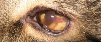
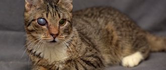
This disease is expressed by damage to the cornea of the eye, which results in the formation of a white scar that can grow and can deprive the pet of vision. Leukoma can be congenital or acquired. With congenital pathology, even in fetal development, infections and viruses of the infected mother are transmitted to the baby, and he is born with a cataract. A cat develops an acquired disease due to injury or the presence of other diseases. Depending on the location of the cat’s thorn, there are three types:
1. Total – the entire surface of the eye is covered. 2. Peripheral - located at the edge of the eye, without affecting the pupil. 3. Central - complete or partial clouding of the pupil.
Murkoshi specialists warn: failure to contact a veterinarian in a timely manner can lead to complete blindness and loss of the eyeball.
Causes of a cloudy film on the eye
Inflammation of the cornea with scar formation occurs when exposed to various pathological factors. In the practice of veterinarians, there are cases of intrauterine leukoma formation in small kittens, and with infections in the pregnant cat itself.
The main causes of eyesore:
- endocrine pathologies of the body (diabetes mellitus);
- mechanical eye injuries;
- poor hygiene when keeping a pet;
- unbalanced diet;
- degenerative processes in the eyeball;
- infectious processes caused by a bacterial, viral or fungal nature.
In addition, there are a number of diseases that affect the eye itself in cats, which ultimately leads to the formation of leukoma. There are a number of main diseases:
- Nuclear sclerosis is a change in the density of the transparent core of the lens with aging, characteristic of older animals. Nuclear sclerosis is caused by excessive compression of old lens fibers in the nucleus and the further development of new ones. The new dense fiber leads to strong dispersion of the light flux.
- Cataracts are a dangerous disease that is diagnosed not only in old cats, but is also observed in young ones. The development of cataracts is mainly provoked by diabetes mellitus and parasitic infestations.
- Glaucoma is a pathological process characterized by pressure inside the eyeball. In this case, the functioning of all functions of the eye is significantly impaired. It increases in size, severe pain occurs, the pet squints its eye, and the cornea becomes cloudy. Edema of the eyelids is also observed, and hemorrhages are observed in the tunica albuginea.
- Conjunctivitis is a pathology that occurs during pathological inflammatory processes in the eye due to a foreign body entering the eye or due to allergic-type reactions. Features of the characteristic inflammation of the conjunctival sac are the accumulation of specific fluid in the area of the outer shell of the eye.
- Uevitis is an inflammation of the vascular part of the eye. This pathology is characterized by the development of severe pain, blepharospasm, and clouding of the fluids in the eyeball. Untimely treatment of uevit leads to the development of adhesions, cataracts, glaucoma and blindness. The main reason for the development of ueviitis in domestic cats has not yet been determined.
- Keratitis is an inflammation of the cornea itself, developing against the background of infectious diseases. More often, keratitis is diagnosed with concomitant systemic intoxications of the body and conjunctivitis.
Symptoms of the disease
Any visually noticeable change in the condition of the eyes signals the need to contact a veterinary clinic. What symptoms indicate the possible presence of cataracts in a cat? Let's look at the most common ones:
1. Covering the ocular surface with a whitish film with a grayish or yellow tint, without black dots. 2. Discharge from the affected area contains pus. 3. The animal is afraid of light and hides in the dark. 4. Deterioration of vision: the cat bumps into objects in a familiar room. 5. Redness and swelling. 6. Nodules may form. 7. Pain when trying to touch the eye.
What cat breeds are at risk?
In 8 out of 10 cases, cataracts occur in older cats - over 8-9 years old. Animals that have crossed this age threshold often face serious illnesses, metabolic disorders, and large amounts of free radicals in the body. The result of these changes is a disruption of the structure of the eye lens.
Has your pet been diagnosed with diabetes? Be careful - this disease increases the risk of cataracts by 20%!
As for the breed predisposition of cats, brachycephalic breeds are most prone to lens clouding:
- British,
- exotic shorthair,
- Persian,
- Scottish Straight and Fold,
- Himalayan
Diagnosis and treatment
In case of bruise or injury to the eye, it is immediately necessary to treat the eye with saline solution or Miramistin and instill drops of Tetracycline. Next, contact your veterinary center immediately. The same advice is also true if you experience any of the above symptoms. To establish an accurate diagnosis, the veterinarian will carry out the following activities:
- measure intraocular pressure; - perform an ophthalmoscopy (examination of the eyeball using special instruments); — will do a general and biochemical analysis.
For serious eye damage, ultrasound is used. If a cat's cataract is detected at the inflammatory stage, treatment is carried out using medications that eliminate foci of infection, discharge and pain. These can be injections of Novocaine, Taufon, Ortofen, Dexamethasone, Levomycetin and others. The main thing is that a scar does not form. In advanced cases, the only way to get rid of cataracts in kittens is through surgery. During the operation, the surgeon removes the cloudy film and replaces it with artificial tissue. Treatment procedures include the following:
- removal of secretions with a swab soaked in a disinfectant solution; - use of anesthetic drops; - placing antibacterial ointment under the lower eyelid; - instillation of drops for itching; - cleansing the eyes with antiseptics.
Self-medication with drugs prescribed for humans is unacceptable. They can slow down the healing process and cause a negative reaction in the pet’s body. Only a veterinarian can prescribe medications for treatment after an accurate diagnosis has been established.
Read more about the dangers of self-medication: Is it possible to treat cats at home?
Therapy
Treating a cat's eyesore requires a comprehensive approach. Of great importance in determining the course of therapy is the diagnosis (statement of the underlying disease that caused leukoma). The stage of the disease and the previous clinical picture matter.
Timely diagnosis and first aid for the cat determine further tactics. In the vast majority of cases, this is conservative therapy with the use of medications.
Severe cases, accompanied by serious symptoms and complications, require surgical intervention. During the operation, the surgeon carefully removes the resulting film, replacing the affected area of the cornea with artificial tissue.
Treatment of an eyesore in a pet with medications includes:
- thorough cleansing of the affected areas of the cornea with special disinfectants;
- use of antibacterial ointment in the lower eyelid area;
- administration of painkillers in the form of drops;
- instillation of drops, widely used in the field of ophthalmology to eliminate symptoms;
- washing the affected eye with antiseptic agents that relieve inflammation and discomfort.
Leukoma can be cured only through complex treatment using evidence-based medicine and traditional methods. It is important to note that infusions and decoctions of medicinal plants to clean the affected eye of a cat can only be used after consultation with the treating veterinarian and in combination with medications. You can use resin from fir trees, celandine mixed with propolis, a decoction of chamomile and calendula.
What is the ocular surface?
The ocular surface is a complex biological structure, including the surface layer of epithelial cells of the cornea, conjunctiva, nasolacrimal outflow system, as well as a secretory apparatus consisting of a complex of multidisciplinary glands and secretory cells that produce components of the tear film, which is an integral part of the ocular surface.
The ocular surface includes the glands of Meibom, the glands of Moll and Zeiss, which produce lipid secretion for the tear film, as well as the secretory glands of Wolfring and Krause, which provide the basal (main) production of fluid for the formation of the aqueous layer of the tear film. In addition, the secretory system of the ocular surface includes Goblet cells, Manz cells, the crypt of Henle and Becher goblet cells, which produce mucin, forming the layer of the same name.
In addition to these structures, the ocular surface includes the hair follicles of the eyelashes and the edges of the eyelids.
Recovery
Recovery and prognosis will depend on the severity of the condition and the effectiveness of treatment. Always follow your veterinarian's instructions carefully after treatment or after surgery. It is essential that you take antibiotics for the entire recommended treatment period, even if the condition begins to improve. Failure to do so may result in relapse of the aggressive disease or loss of vision.
Corneal ulcers and keratitis typically begin to heal within three to five days after treatment. Some ulcers caused by infection take longer to heal. Your veterinarian will advise you on your cat's signs.
For cataracts and glaucoma, your veterinarian may schedule follow-up visits as needed to monitor the condition. If cloudy under eyes do not improve or seem to get worse despite treatment, contact your veterinarian immediately.
General information
The cornea is a relatively dense, transparent layer that protects the front, exposed surface of the eyeball. Both the shape of the cornea (it should be as flat, smooth, spherical as possible) and its transparency are of key importance, since any optical interference - turbidity, inhomogeneous inclusions, scars, etc. - distorts the normal refraction of light and/or reduces its intensity on the way to the retina inevitably affects the quality of the final visual image. Unlike some other inflammatory processes of the eye (blepharitis, conjunctivitis), which may not significantly affect visual acuity, with keratitis there is almost always a decrease in visual function. That is why it is extremely important to begin treatment as soon as possible, before irreversible scar changes form on the main optical axis of the eye.
Treatment of eye injury
Minor and uncomplicated injuries are treated on an outpatient basis, while more serious eye injuries are treated in a hospital setting.
In case of injury to the eye membranes, surgical procedures are performed. For minor eye injuries, primary treatment of the wound is performed, and in more serious cases, removal of foreign bodies from the eye cavity, plastic surgery of the eye and restoration of its structures.
"MedicCity" provides assistance to patients with any ophthalmological diseases. The clinic's ophthalmologists are proficient in all modern methods of diagnosis and treatment in ophthalmology and use high-precision equipment from global European manufacturers in their work. Trust us and we will take care of your vision!
Viral keratitis
Keratitis caused by viruses is often accompanied by a blistering rash. The pathogenic agent can be almost any aggressive virus (measles, chickenpox, adenoviruses), but most often it is the herpes virus. As is known, over 95% of the population is infected with herpes infection, and in most cases, herpes occurs latently, in an asymptomatic form, and is activated only when immune resources are weakened, general exhaustion of the body and other unfavorable conditions. In this case, inflammation of the cornea is usually preceded by typical herpetic symptoms - rashes on the lips or other mucous membranes. With herpetic keratitis, as a rule, swelling and the appearance of fuzzy, vague infiltrates predominate.
Types and types of keratitis
In publications you can often find a division of keratitis into exogenous (caused by external influences) and endogenous (caused by internal factors present in the body). However, such a classification seems too general and not entirely successful; It is unclear, for example, to which class allergic keratitis associated with wearing contact lenses should be classified: on the one hand, the allergic reaction is caused by internal immune disorders, on the other, by an external, artificial object.
According to the dynamics of development and type of course, acute and chronic keratitis are distinguished; as a variant of the second type, recurrent keratitis is sometimes considered separately.
The depth of spread of the inflammatory process is essential: superficial keratitis is much less dangerous than deep keratitis, in which the inner corneal layers are scarred.
According to the degree of severity, keratitis is usually divided into mild, moderate and severe; according to the localization of inflammation - central and peripheral.
However, the most convenient and intuitive is the etiological classification, where keratitis is grouped according to the direct causes of the inflammatory process.
At what age does dry eye syndrome most often appear?
Dry eye syndrome can be detected in patients at almost any age. But most often it is diagnosed in patients over 40-50 years old. However, due to environmental changes, the widespread use of technology at work and at home, and the increasing use of contact lenses, the age of patients with detectable dry eye syndrome is becoming younger.
You need to understand! If everyday life is associated with the constant presence of risks or taking medications, the side effects of which disrupt the secretory function of the glands, then regular monitoring by an ophthalmologist is necessary in order to timely correct and take the necessary measures.
Bacterial keratitis
Keratitis can be caused by a variety of pathogenic microorganisms, primarily cocci (Staphylococcus aureus, Streptococcus, Gonococcus) and Pseudomonas aeruginosa. A rare and very dangerous infection, even resulting in blindness, is acanthamoeba infection (named after the causative agent Acanthamoeba), which can exist, in particular, in the gap between the contact lens and the surface of the cornea. With the development of the so-called creeping corneal ulcers caused by gonococcal, tuberculosis, syphilitic and other bacterial infections, the risk of rapid and irreversible loss of vision is also very high.
First aid for eye injury
To properly assist a person with an eye injury, you must adhere to the following rules:
- if small foreign bodies get into the conjunctiva, you can try to wash them with running water;
- You should not try to independently remove a foreign body stuck in the deep membranes of the eye;
- the injured eye must be covered with a clean gauze bandage (cotton wool cannot be used!);
- in case of severe pain, it is recommended to take a painkiller;
- It is necessary to consult an ophthalmologist as soon as possible.