Signs Diagnosis Treatment The diaphragm is the muscular partition between the chest and abdominal cavity. Performs an important respiratory and barrier function. A diaphragmatic hernia occurs when the integrity of the diaphragm is disrupted or ruptured. In this case, the abdominal organs are displaced into the chest. Most often, a diaphragmatic hernia occurs due to trauma and is combined with multiple injuries (a cat gets hit by a car, falls from a window).
There are 2 types of diaphragmatic hernia:
- traumatic – rupture of the diaphragm due to a strong blow or injury;
- congenital - a cat is born with a defect (the most common is peritoneo-pericardial diaphragmatic hernia, in this case the abdominal organs are displaced into the pericardial cavity through a hole in the diaphragm).
Diaphragm and diaphragmatic hernia: how they are related
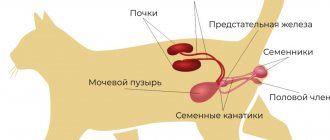
The diaphragm is a powerful inspirator; without it, normal operation of the abdominal press, as well as stable flow of blood and lymph through the main vessels of the abdominal cavity is impossible. The muscular-tendon septum is attached from the inside to the ribs and has the shape of a dome, the convex part of which “looks” into the chest cavity. The diaphragm itself contains openings for the esophagus, aorta, and vein (caudal cava).
Diaphragmatic hernia develops in cats as a pathological condition due to various reasons. But in this case, damage to the “film” of varying intensity occurs, in which organs from the abdominal cavity begin to shift upward, causing pinching of the lungs, heart and large vessels.
Aperture functions:
- participation in breathing;
- support for organs;
- natural partition;
- providing lymphatic drainage;
- participation in digestion.
A hernia most often appears when the animal is severely injured (road accident, fall from a height) or due to an impact. Along with the hernia, the cat has multiple injuries to the bones and soft tissues, so it is necessary to take it to the RosVet EC, where the operating team will take care of it.
Veterinary clinic of Dr. Shubin
Description and reasons
Diaphragmatic hernia is a violation of the integrity of the diaphragm, accompanied by displacement of the abdominal organs into the chest cavity. The diaphragm is a muscle-tendon plate that separates the abdominal cavity from the thoracic cavity. Mammals are the only ones in which complete muscular separation of these cavities is determined (± crocodiles). The formation of the diaphragm during evolution has dramatically improved lung function. The esophagus, posterior vena cava and aorta pass through this plate.
Drawing. Diaphragm of a dog, view from the abdominal cavity. Source. Guide to the dissection of the dog, Seventh Edition.
Diaphragmatic hernia can be congenital or acquired. The congenital form of diaphragmatic hernia is rarely diagnosed in a veterinary clinic, because Death with this type of hernia in an animal occurs either at birth or in the neonatal period (immediately after birth). Acquired form of diaphragmatic hernia - develops secondary to any mechanical impact; It is with this disease that we have to work and further we will talk about it. So, a traumatic hernia develops as a result of any impact of mechanical factors on the body, which is often noted in an accident, a fall from a height, and bites by other, larger animals.
Depending on the time of occurrence, diaphragmatic hernia is divided into acute and chronic. The bottom line is that the development of this type of hernia does not always lead to the death of the animal (only 15% of animals die before diagnosis), and cases of diagnosis in animals in a period remote from the injury are quite common.
The movement of abdominal organs through the diaphragm leads to compression of the lungs, which is accompanied by visible manifestations of respiratory failure. At the beginning of diaphragm rupture, these signs are most pronounced, then they may decrease somewhat. However, it is difficult to recognize the presence of a hernia only by external signs of difficulty breathing. Thus, during routine sterilization of cats, there are frequent cases of difficulty breathing and detection of a diaphragmatic hernia in an emergency (radiographic examination already under anesthesia).
Clinical signs and diagnosis
Diaphragmatic hernia can develop in both dogs and cats. In many animals (15%–25%), a hernia is diagnosed within the first week after any traumatic exposure. Due to the fact that this type of hernia develops against the background of various injuries, various concomitant lesions may be detected in the animal (eg severe blood loss, shock, bone fractures). In the chronic course of a diaphragmatic hernia, signs of the disease can be expressed in various types of respiratory disorders (eg shortness of breath, exercise intolerance) or gastrointestinal tract (eg lack of appetite, vomiting, diarrhea, weight loss, pain after eating). As mentioned above, many animals with a chronic form of diaphragmatic hernia do not exhibit dyspnea at the time of examination.
When examining the animal, the veterinarian may identify some signs that suggest the presence of a hernia (eg breathing problems, decreased abdominal volume), but these only serve as a reason for further diagnosis (usually a radiographic examination).
The final diagnosis of a diaphragmatic hernia is usually determined by radiographic examination (X-ray); in rare cases, the use of contrast and ultrasound diagnostics (ultrasound) may be used. Diagnosing most cases of clinically significant forms of diaphragmatic hernia using x-rays usually does not cause difficulties.
Preoperative preparation
An operation to correct a diaphragmatic hernia may require some preparation; this is aimed at reducing the probable anesthetic mortality of the animal and making certain procedures easier to carry out. In most cases, an intravenous catheter is installed and infusion therapy is administered. In case of severe breathing difficulties, the animal can be provided with oxygen, and its placement is carried out taking into account the facilitation of respiratory movements. If concomitant diseases are suspected, the veterinarian may recommend various examination methods (eg blood tests, ultrasound, x-rays). Immediately before the operation, the animal is given an antibiotic.
Anesthesia
Carrying out anesthesia in patients with diaphragmatic hernia is associated with a high risk of complications, including fatal ones. Total intravenous anesthesia (injection of drugs into a vein) is used as the basis of anesthesia, because artificial ventilation is used intraoperatively and this greatly complicates the administration of inhalation anesthesia. An animal with a diaphragmatic hernia is subject to mandatory intubation - a special tube is inserted into the trachea under anesthesia, through which the breathing function is maintained.
Anesthetic mortality during surgical correction of diaphragmatic hernia in our clinic tends to zero, but be that as it may, employees warn every animal owner that any anesthesia can lead to the death of the animal.
Drawing. A ventilator used in a veterinary clinic to maintain breathing in animals during the correction of a diaphragmatic hernia. This device provides excellent intraoperative ventilation and reduces the risk of lung injury due to excess pressure of the inhaled mixture.
Surgical correction
Diaphragmatic hernia is a primarily surgical disease; its correction is achievable only by using various methods to restore its integrity. Conservative treatment (eg intravenous and antibacterial therapy) is aimed only at facilitating surgical intervention. The doctor may slightly delay the operation for drug correction of various complications, however, the operation should be performed immediately after the animal has been stabilized, this improves the prognosis for a favorable outcome.
A patient with a diaphragmatic hernia is placed in a special groove on the back, the incision is usually made along the midline of the abdominal wall from the xiphoid process of the chest half or two-thirds of its length towards the tail. The abdominal organs are shifted to their proper physiological position, which frees up the chest cavity. After achieving the correct anatomical location of the internal organs, the defect of the diaphragm itself is assessed and approximating sutures are placed on its edges. After which, excess air is removed from the chest cavity. In chronic cases of diaphragmatic hernia, various defects and adhesions between internal organs may develop, and they are also subjected to various types of surgical procedures. When correcting a diaphragmatic hernia, non-absorbable sutures (nylon, etc.) are often used.
Postoperative care
The immediate postoperative period is very important in terms of the animal’s recovery; staff “takes” the animal out of the operating room only after spontaneous breathing has been restored and in a stable condition. After the operation, antibiotics and painkillers are administered for three to four days. The optimal way of postoperative care is to keep the animal in a 24-hour hospital, but at the moment we cannot provide this type of service.
Sutures on the abdominal wall are subject to mandatory protection for a period of 14 days; for this, either a protective blanket (optimally) or an Elizabethan collar is used. Protecting the wound prevents the animal itself from affecting the stitches, which reduces the risks of various complications. During this period, the wound itself is monitored for the development of swelling, redness, etc., at the end of this period, the stitches are removed in a veterinary clinic and there is no need for protection. If there is a change in the general condition of the animal, the veterinarian must be immediately informed for a competent assessment and correction of any complications.
Possible complications and prognosis
Postoperative prognoses for animals with diaphragmatic hernia have improved significantly over the past few decades. Thus, in the 1980s, only 52% of cats and dogs underwent surgical correction; recently, the survival rate for diaphragmatic hernia was 82%-89%. Survival data differ slightly between cats and dogs, and between acute and chronic forms of diaphragmatic hernia. However, there continues to be a generally accepted opinion that survival rate for chronic hernia is somewhat lower than for acute hernia. The main reasons for the unfavorable outcome of hernia correction surgery are considered to be deficiencies in anesthesia and artificial ventilation.
Various complications develop in half of patients after surgery to correct diaphragmatic hernia. Fatal complications can develop from both the thoracic and abdominal organs. Also, complications can develop immediately after surgery or at a somewhat distant period of time. Soon after surgery (the first day), death may develop for the following reasons: accumulation of blood, air and fluid in the chest cavity; pulmonary edema; shock, heart rhythm disturbance. In the long term, death may occur secondary to rupture, torsion, or obstruction of the gastrointestinal tract or diseases unrelated to hernias. Recurrences of a hernia after proper correction are quite rare, but they can still amount to 4%-5%. Rarer complications may also occur and should be discussed with your veterinarian.
Conclusion
Be that as it may, with surgical correction of diaphragmatic hernia, a fairly high recovery rate is observed in cats and dogs. The staff of the veterinary clinic have accumulated enough successful operations, and in our opinion, the only way to restore the health of the animal is to suturing the diaphragm defect.
Screening image of a kitten admitted to the veterinary clinic with severe shortness of breath; the x-ray is not of the best quality, but it clearly shows signs of a diaphragmatic hernia. Due to the serious condition of the animal, the veterinary clinic staff did not conduct a detailed X-ray examination, but decided to undergo surgery.
The same kitten the day after surgery in the veterinary clinic. The veterinary clinic staff, under anesthesia, returned the organs to their normal position and sutured the diaphragm defect. While in the hospital at the veterinary clinic, a “butt with a handle” was stuck to the kitten due to its catastrophic thinness, long tail and some character traits.
The same kitten, 14 days after surgery, allows doctors to remove the stitches.
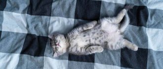
He looks serious and looks confidently into the future.
July 2016
Veterinary clinic of Dr. Shubin, Balakovo.
Types and characteristics of diaphragmatic hernias
After the examination, the veterinarian determines the cause of the damage; this is what is important to do in order to predict the course of the operation in the future. A defect in the diaphragm appears after an injury; these are the most common cases of cat owners contacting the RosVet VC.
Mechanical factors:
- closed (bumps, falls, road accidents, sharp increase in intra-abdominal pressure);
- open (when, together with the diaphragm, the integrity of the surrounding tissues and bones is disrupted).
Clinical signs of damage are very different and depend on the cause, on which organs and tissues are injured. The respiratory and digestive systems are most often affected.
With a congenital diaphragmatic hernia, there are:
- pleuroperitoneal, which are rare and kittens with similar anomalies die in the first week after birth;
- pericardio-pleuroperitoneal, the most commonly diagnosed injuries in the practice of veterinary surgeons, Persian cats are predisposed to them;
- sliding, develop with weakness of the esophageal-diaphragmatic ligament, while the stomach and esophagus, having lost support, move into the mediastinum. A hernial sac is formed from a fold of the peritoneum and, as a result, the sphincter between the stomach and esophagus does not close well (reflux esophagitis);
- paraesphageal, the fundus of the stomach, omentum and part of the intestine are moved through the dilated esophageal opening into the chest cavity with simultaneous fixation of the cardiac section. The main difference is the possibility of strangulation, pain, nausea and vomiting. In the absence of quick help, the strangulated tissues quickly die, which is irreversible.
Traumatic injuries can cause pneumothorax (air in the chest cavity) or hemothorax (collection of blood). As a result, breathing is impaired until critical situations arise when the cat needs help immediately.
Features of the disease
Animal hernias are similar in structure to such formations in humans. They consist of a hernial orifice and a sac with contents (part or all of the peritoneal organ). In young animals, the hernia most often involves part of the stomach or intestines. But the defect can also affect other organs, such as the bladder.
The main danger of a hernia is that, under certain circumstances, an organ caught in the hernial sac is strangulated. As a result, its blood supply is disrupted, which can lead to necrosis, that is, death. Constipation, severe coughing, and abdominal wall injuries contribute to the development of strangulation. For example, a kitten's hernia may be strangulated after an injury caused by an aggressive animal.
What symptoms should alert the owner?
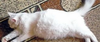
If the organs of the abdominal cavity are shifted through the hole into the chest cavity, then compression of the lungs inevitably occurs, and also the organs themselves, pinched in the diaphragmatic hernia, have their blood circulation disrupted.
The following signs should alert a cat owner:
- obvious breathing problems;
- a superficial, shallow sigh;
- the cat deliberately extends its neck and head to improve air passage.
The liver, stomach and intestines “stuck together” cease to function normally, which visually may look like signs of gastrointestinal disorders or liver failure.
Causes of hernia formation
The main factors leading to the appearance of this pathology include:
- heredity. If one of the kitten’s parents had a congenital hernia, the likelihood of detecting a similar pathology in the offspring increases significantly;
- intestinal problems (constipation, flatulence);
- cutting the umbilical cord too short or the cat biting it off.
Various injuries can also lead to the formation of a hernia. Most often, the cause of its appearance is falls, bruises, bites from dogs or other animals.
What kind of research is carried out at the RosVet EC?
Diagnostic measures necessarily include radiography. If there is no clear line of the diaphragm on the image, the veterinarian always suspects a diaphragmatic hernia. Visualization of the internal organs in the thoracic region, which anatomically should be located in the abdominal cavity, only confirms the diagnosis.
Contrast studies are necessary if there is pleural effusion. Peritoneography is performed to confirm rupture of the diaphragm; if there is damage, then the contrast agent will be in the chest cavity. Ultrasound is prescribed as an auxiliary study.
Prevention
The main measure to prevent the formation of a hernia in kittens is a special massage . Only a qualified veterinarian can show you how to do it correctly. Regularly performing such a massage will be useful for any kittens, but it is especially necessary for those whose parents suffered from hernias. It can significantly reduce the likelihood of deformities in the abdominal wall. In some cases, with the help of massage it is even possible to eliminate small hernias in the initial stages of formation.
It is also recommended to undergo regular examinations by a veterinarian. It must be taken into account that an animal that has been diagnosed with a congenital hernia cannot be used for breeding.
What to do: what treatment is provided
There is no drug treatment for diaphragmatic hernia; drugs are prescribed as auxiliary ones, but the main therapy is only surgical. Moreover, the faster the cat owner delivers the pet to the clinic, the higher the chance of a successful outcome.
The surgical operation consists of suturing the hernial opening in the diaphragm. If stabilization of the animal before the intervention is impossible, then an emergency operation is performed using a ventilator and the cat’s further stay in the intensive care unit and hospital of the veterinary clinic.
If your cat has suffered serious injuries (road accident, bruises from a fall, fractures, etc.), it is necessary to urgently deliver it to the RosVet EC. You can inform about your arrival by phone: + 7 (495) 256-11-11. Also, with a congenital pathology, the owner may notice signs of breathing problems, which will also be a reason to come to the clinic or call a doctor at home.