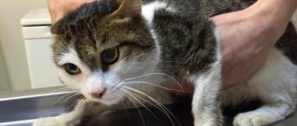
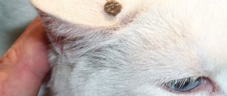
Lipoma (wen, steatoma, lipid cyst) is a benign limited tumor of the skin formed from adipose tissue. All species of mammals are prone to it, regardless of gender and age.
Dogs and ferrets are more susceptible to developing lumps on their bodies than cats because their lipid metabolism is poorer than that of felines. This pathology is not an infection, so the possibility of infection of a healthy animal from a sick one is excluded, but requires the intervention of a veterinarian.
The number of wen can reach ten or more (lipomatosis), the size depends on the time of appearance and location. Size varies from pea-sized to large growths.
There are giant seals with a hemispherical flattened configuration. Sometimes they are pear-shaped, located on a thin stalk.
Why are fatty cysts dangerous?
The location can be any, but more often it is the subcutaneous tissue of the back, neck, chest, and limbs. A small growth on the body, in the singular, does not cause much harm to the pet, but as the growth increases, discomfort and a deterioration in the quality of life are possible.
Located on the neck, the lump can compress large blood vessels that supply the brain, which leads to oxygen starvation of the tissues.
Large wen on the limbs or armpits cause inconvenience when walking, which is why the animal moves little and refuses to walk.
As the growth grows, the skin stretches, causing itching. Trying to get rid of it, the pet licks the surface, chews out tissue, as a result of which pyogenic bacteria enter the wound, causing an inflammatory process.
In females, a lipoma can form in the mammary glands. The neoplasm is injured and is also prone to degeneration into cancer.
Often fatty cysts appear inside the abdominal cavity, causing functional disruption of organs, their displacement and compression.
Disease prognosis
The prognosis of the disease, subject to surgical intervention, is favorable, especially if the size of the wen is small and the cat is in good physical condition. Relapses of the disease are rare. Almost all cases of relapse relate to infiltrating variants of the disease. Recently, veterinarians have been practicing radiotherapy for such forms of the disease, which has significantly increased the percentage of cases of complete recovery.
Lipoma or wen are diseases for which self-medication is unacceptable. At best it will be ineffective, and at worst it will be harmful. Therefore, the only possible way out in such a situation is to visit a veterinary clinic.
How to recognize a wen in an animal
Steatoma differs from other neoplasias in its slow growth and the presence of clear contours. It is mobile and painless. Upon palpation, it is discovered that the seal is not fused to the skin and deep tissues and has a soft dough-like consistency.
Often, palpation reveals fluctuation (a feeling of oscillation), which can be confused with an abscess. If the neoplasm is not burdened by pathological changes, then no subjective sensations are observed in the animal.
However, large growths can lead to changes in the functioning of internal organs and the occurrence of diseases. Sometimes dense areas alternate with soft ones, which also leads to incorrect diagnosis.
An important sign of fat formation is the presence of lobules. The skin in this area does not change color, sometimes it can acquire a yellowish-brown tint.
Clinical signs of lipoma
The location of the cones varies, but if they are located in a vital area, they can pose a serious threat to the health of the animal.
The appearance in the neck area makes breathing difficult, which causes hypoxia to develop. The animal shows signs of depression of the cerebral cortex.
It is noted that:
- lethargic sleepy state
- lethargy
- nausea, vomiting
- urinary and fecal incontinence
If there is a lipoma in the head area, neurological disorders may occur due to compression of the vessels supplying the brain.
The animal is observed:
- change of consciousness
- impaired movement coordination
- convulsions
Severe brain hypoxia leads to the death of the pet.
Lipomas on the paws or armpits lead to lameness and changes in gait.
Mastopathy
Mastopathy can be classified as benign subcutaneous formations that can occur in the mammary gland of a cat and bring it discomfort, even pain. Subcutaneous lumps appear in the mammary gland. Although it is absolutely possible that the mammary gland may enlarge for another reason. For example, the mammary gland can enlarge during pregnancy. It is noteworthy that during false pregnancy the mammary gland can not only swell, but also secrete a little milk. But within a short time everything should return to normal.
Mastopathy is a permanent pathological transformation in the tissues of the mammary gland. They observe the appearance of neoplasms, as well as other abnormal processes. Formations in the form of lumps in the mammary gland can be felt during palpation. They are soft and slightly elastic in nature. Mastopathy in dogs cannot be classified as an oncological disease, but it can be a harbinger of such a disease. Cats over 6-7 years of age are most prone to mastopathy.
There are two forms of mastopathy in cats:
- Diffuse mastopathy. The first sign of this form is the appearance of pain in the mammary gland shortly before estrus. Upon palpation in the chest, you can feel a formation similar to a bag of shot. The diffuse form may be a precursor to the fibrocystic form.
- Fibrocystic mastopathy. In the mammary gland, compacted painful nodules form, which tend to grow. Lumps on the chest can be either single or multiple, but are always easily identified by palpation. This diagnosis is most often made in cats over 6 years of age.
Fibrinous cystic mastopathy of severe generalized form. All mammary glands are involved in the process.
To recognize mastopathy in time, you need to take a close look at the following symptoms:
- Noticeable enlargement of the mammary glands. When palpated, you may notice graininess, lobulation or stringiness.
- Various pathological discharges from the gland may appear: blood, ichor or colostrum.
- The cat may be in pain. At the same time, she will try to lick the glands and will be restless. It is difficult to notice any deviations from the norm, because the formation can remain the same size for a long time. The dimensions of the nodule can change only in the case of false or normal pregnancy.
- If treatment is not started on time, the dog’s general condition worsens sharply. He may refuse to eat, but increase his need to drink. Apathy is clearly visible in behavior. Nearby lymph nodes may grow noticeably. Ulcers and suppurations may appear. With further inaction, the process spreads to adjacent tissues. In this case, the animal’s skin in this area becomes too hot and may even lose hair.
It is very important to recognize the stage at which mastopathy is located. In the early stages, the use of homeopathic medicines and diet is sufficient. The diffuse form can be cured with hormonal drugs and antibiotics.
The difference between a benign tumor and cancer
A malignant lump is usually lumpy and has indistinct edges, unlike a lipoma, which appears as a smooth, spherical formation. Cancerous tissue grows quickly and is prone to ulceration, while wen develops slowly (if it does not grow into deep tissues) and is injured only under external influence.
A fatty cyst is usually soft, while a cancer lump is always hard and elastic.
There are other types of growths that require special attention and treatment to avoid serious complications.
It can be:
- papilloma (wart)
- Tumor
- abscess
- cyst
- hematoma
- mycetoma
- sporotrichosis
Types of lipomas
Wen in cats can be single in appearance, or can be multiple in nature, especially in the presence of concomitant diseases (deviations in the functioning of the endocrine system). This multiple formation of wen is called lipomatosis.
According to the nature of germination, a lipoma can be simple or infiltrating:
- A simple lipoma has clear boundaries and, as it grows, does not affect the surrounding muscles, so it can easily be surgically removed.
- Infiltrating lipoma grows into the thickness of the muscles and vascular bundles and does not have a clear boundary. It is this type of neoplasm that tends to degenerate.
Diagnosis of the disease
Fats that tend to grow into blood vessels and deep tissues (infiltrative lipomas) are often confused with cancer, so they must be examined carefully.
To determine the nature of the neoplasm, a biopsy (tissue sampling) is performed. In case of infiltrative compaction, computed tomography (CT) is prescribed to determine the extent of tumor damage to blood vessels and muscles.
Histological analysis
Lipoma consists of lobules of adipose tissue separated by connective tissue septa. If there are few blood vessels, then it is dense and damage does not lead to heavy bleeding.
Any neoplasm on the animal’s body must be examined, since there is a risk of degeneration of a benign tumor into a malignant one (liposarcoma).
Rehabilitation
The duration of the rehabilitation period after surgery depends on the nature of the lipoma. Restoring your pet’s health after removal of large infiltrative lipomas can take a long time. During this period, the cat needs special care.
It is very important to monitor the condition of the seams. At the slightest suspicion of inflammation, the animal should be taken to a veterinarian. After the operation, you need to put a special protective collar on the cat so that the pet does not lick the wound.
It is also necessary to ensure that the postoperative bandage fits snugly to the body. If the bandages are loose or dirty, then it is necessary to bandage them.
After removing a wen on a cat’s abdomen, a special bandage should be used. This will help protect the postoperative suture from infection.
Treatment of wen in animals
If you find a lump on your pet's body, do not try to remove it yourself, puncture it, or squeeze out the contents. Such manipulations can cause bleeding, inflammation, and degeneration into a malignant form.
A lipoma in an animal requires examination and consultation with a veterinarian! After examination and laboratory diagnostics, the specialist decides on the treatment method.
If the appearance of neoplasia is caused by consuming large amounts of lipids, then changing the diet may reduce the tumor, but over time, growth may resume.
Small lipomas are observed and, as they grow, removed under local anesthesia at home, which greatly facilitates the rehabilitation period. Large formations must be removed immediately, as they can cause inflammation or lead to disruption of the functions of vital organs.
Spontaneous disappearance is a rare occurrence.
Causes and symptoms of lumps
A cat's lump under the skin can be malignant or benign. In the first case, the pet should be prescribed treatment as soon as possible, but it may not bring a positive result.
But the lump can also be a benign neoplasm. In this case, there is no need to panic, but it is better to show the cat to the veterinarian as soon as possible so that he can do the necessary research.
If a cat has a lump, this does not mean that the pet is experiencing pain or stress - often the lumps are painless.
Lumps can be very painful - most often this indicates some kind of disease. Even if the lump is not painful and benign, even this can significantly worsen the animal’s life. It is especially dangerous if the lump occurs on the back: this can lead to blockage of blood vessels, blood and oxygen do not reach the organs, and this condition can result in paralysis of the limbs or death.
Establishing diagnosis
Due to the fact that lipomas do not cause pain, owners often do not pay attention to them. However, after detecting a formation in a cat, it is important to consult a doctor to determine the correct diagnosis. This is explained by the fact that only in the clinic will they be able to determine whether it is a wen or other types of malignant tumor. And also prevent the growth of the tumor. Wen is not an infectious disease and therefore cannot be transmitted to people or other pets.
The veterinarian carries out a number of preliminary measures:
- signs of a wen in a cat are determined by external examination;
- X-ray, ultrasound; puncture biopsy.
© shutterstock
When palpating a cat, single wen can be easily felt. They are soft and easy to move. An x-ray will determine the presence of metastases. In cases where wen is located in hard-to-reach areas, ultrasound will help determine the need for surgery. Using a biopsy, the type of cells that make up an organ or formation in it is determined. A puncture biopsy will clarify the nature of the tumor: benign or malignant. Cytological examination is carried out before surgery.
Clinic
Older cats are most often affected. These animals suffer much less often than their canine counterparts. If any formation appears on the skin of an animal, you should immediately seek qualified medical help to exclude a cancerous tumor and carry out treatment in a timely manner.
The symptoms are always the same; swelling appears on some part of the body. She can remain in this position for a long period of time. Over time it grows, the sizes may vary. The pet's general condition does not suffer, it may cause discomfort when walking or lying down, depending on the location of the wen.
Veterinarians, having carried out an examination and diagnosis, remove the tumor, followed by a histological examination of the cells.
Is the operation effective?
Can lipomas reappear after surgery? In 70% of cases, no recurrence of tumors is observed. Veterinary experts have found that removing the wen promotes the overall health of the body and prolongs the life of the pet by several years.
The situation becomes somewhat more complicated if the cat suffers from multiple lipomas (lipomatosis). In this case, after the operation it is necessary to review the pet’s diet. You may need to follow a lifelong fat-restricted diet, as well as take medications that normalize lipid metabolism. This will help prevent the recurrence of lipomas.


