Alas, pets are not only happiness, but also a great responsibility, since it is people who determine how long and carefree a pet will live. He cannot talk about his poor health, so the task of a responsible owner is to ensure that the kitten is well-groomed, fed, and feels good. It is important to pay attention to external signs of disease.
The article will tell you why a cat’s eyes are half covered with a film, what are the causes of the disease and methods of its treatment.
Short description
Unfortunately, a disease in which part of a cat's eyelid is covered with a white, opaque film is common. The film bears the name of the third century and is of great importance. It removes foreign particles and dust, and also distributes tear fluid. The third eyelid (conjunctival fold or nictitating membrane) is located at the inner corner of the eye. It is almost impossible to notice it in a healthy animal. If the cat is sick, the membrane becomes inflamed and turns white.
Functions of the third eyelid
A cat's third eyelid is a small, light-colored (sometimes pigmented) fold that is located at the corner of a cat's eye. It performs an important function, since it helps moisturize the cornea. The third eyelid helps protect the eye from dryness, dust and debris.
The third eyelid can also be seen in completely healthy cats. In a normal state, it should not be visible all the time: only when the cat tilts its head, and sometimes the nictitating membrane opens during the animal’s sleep. If half of a cat's eye is constantly covered with a thin fold, this is the first sign that indicates a possible disease in the animal.
When the third eyelid is damaged or falls out, the cornea often dries out, which can cause conjunctivitis. A prolapsed nictitating membrane is very vulnerable to various infections. Its infection by viruses can lead to complete blindness of the animal, and without timely treatment, the infection can spread to the brain.
Main reasons
The reasons for the appearance of a white film on a cat’s eyes can be varied:
- eye diseases such as conjunctivitis;
- mechanical impact;
- getting foreign objects into the eyes;
- parasites (internal and external);
- neoplasms (adenomas);
- allergy.
Let's look at some of them in more detail.
Infection
The action of the main contagion is combined with the influence of secondary microflora:
- rhinotracheitis;
- calcivirosis;
- panleukopenia;
- chlamydia.
Congenital anomalies
It is believed that some cat breeds are genetically predisposed to eye diseases. At risk, for example, are Persian cats and other brachycephalic breeds.
Invasion
Eye inflammation is caused by the following pathogens:
- protozoa;
- toxoplasmosis;
- helminthiases.
Permanent pathologies
These include pathologies such as hepatitis, pancreatitis, diabetes mellitus and other chronic diseases. They weaken the immune system, making the cat defenseless against the microflora that causes conjunctiva.
Hypersensitivity
Plant pollen, perfumes and accessories can be irritants in the formation of conjunctivitis, which is accompanied by the appearance of a transparent film on the cat's eye.
Why does a cat have a film on its eyes?
There are several reasons for the appearance of this neoplasm in the eyes of a pet. Firstly, it could be a speck that got into the eye and caused swelling, as well as triggering an inflammatory process. Secondly, the film could appear due to diseases of an infectious nature. If in this case no measures are taken and the animal is not helped, the cat may go blind.
Also, the cause of eye film can be parasites, diseases of internal organs, in particular intestinal and stomach diseases, conjunctivitis, bacterial infections, mechanical damage to the eyes, strabismus, cataracts and diseases of the vascular system that nourishes the organs of vision.
External manifestation
Third eyelid disease can be diagnosed not only by changes in the pet’s appearance, but also by changes in behavior. Most likely, the cat is hiding from bright lighting, rubbing its face with its paws, and especially its eyes, blinking and squinting frequently due to noticeable discomfort. You can also see profuse lacrimation and even the presence of pus. If treatment is not carried out in time and you do not consult a veterinarian, there is a high probability of vision loss.
You can also notice the third eyelid by the presence of the following inflammations:
- adenoma;
- cartilage malformations;
- protrusion of the nictitating membrane.
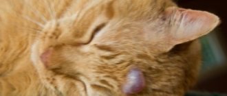
Adenoma
This is a pink tumor that is located near the bridge of the nose. It is benign and resembles a bean in shape. The main causes are injury or infection. It is easy to detect; the eye stops closing. If not treated promptly, serious complications can occur.
Cartilage malformations
Defective cartilage is deformed during the growth phase, as a result of which the nictitating film begins to become inflamed. It is one of the congenital anomalies.
Protrusion of the nictitating membrane
If the third eyelid appears in only one eye, the causes of the pathology may be as follows:
- foreign objects under the film;
- injuries to the facial nerves, cornea or cartilage;
- accretion of the membrane to the eyeball.
If the third eyelid appears in both eyes, the following reasons for its occurrence are distinguished:
- infectious conjunctivitis;
- helminthiases.
Causes and symptoms of eye diseases in cats
The most common diseases in cats are:
Inflammation of the eyelids
. With phlegmous inflammation, the eyelid swells and purulent mucus flows from the eye. With normal inflammation, the cat begins to scratch its eyes. The eyelids become red and thickened. The disease appears due to vitamin deficiency, eczema. Phlegmous inflammation develops after strong calculations and deep wounds.
Bruises and wounds
. A cat can receive these injuries from a fall or after a fight. The wound can be superficial, deep or through. The main symptom is severe swelling, redness and even bleeding from the eyes.
Turn of the century
. The skin turns inward. This causes a strong inflammatory process. Volvulus can be caused by a foreign body getting into the cat's eye or exposure to chemicals. Without timely assistance, the cat may develop conjunctivitis or keratitis. In advanced cases, an ulcer will appear on the cornea.
Symptoms of the disease include lacrimation and photophobia. The eyelid begins to swell, changing its appearance.
You can recognize the listed eye diseases in cats from photos.
Types of conjunctivitis
Conjunctivitis is considered the most common disease among cats. It has the following varieties:
- Purulent.
- Allergic.
- Acute catarrhal.
- Follicular.
With purulent conjunctivitis, the general condition of the cat worsens. The animal's body temperature rises, and copious pus begins to discharge from the eyes. Diarrhea and vomiting may occur.
Allergic conjunctivitis
in a cat it causes contact with an allergen. At first, the discharge from the eyes appears clear. If left untreated, they become purulent.
Acute catarrhal conjunctivitis is always accompanied by redness of the eyes and severe swelling. Cats experience pain, tears flow, and serous-mucous discharge occurs. The main reason is injury and lack of vitamin A in the body.
Follicular conjunctivitis is characterized by inflammation of the lymphatic follicles. They are on the inside. The disease is chronic and requires long-term treatment. Surgery is possible.
The photo clearly shows all types of conjunctivitis.
Types of keratitis
Keratitis is a disease of the cornea of the eyes. The most common types are:
- Superficial purulent.
- Vascular superficial.
- Purulent deep.
With superficial keratitis, the upper (epithelial) layer of the cornea becomes inflamed. The cat is in pain and afraid of light. The cornea takes on a gray color and swelling develops. This type of illness usually occurs due to injury.
With vascular keratitis, capillaries grow into the upper layer of the cornea, causing clouding of the eyes.
Purulent deep keratitis is a very serious disease caused by microbes that have penetrated the stroma of the cat’s cornea. The cat begins to be afraid of light and scratches its eyes continuously. The cornea takes on a yellowish tint. Capillaries begin to grow into the cornea. The cause of the disease is considered to be injury and infection. The cat will be sick for a long time.
Corneal ulcer
To this disease. Causes infections that develop after deep wounds. This may also be a complication after suffering purulent keratitis. There are 2 types of ulcers: perforated and purulent. The main symptom is severe pain. Therefore, the animal is always restless.
When a perforated ulcer appears, purulent discharge from the eyes is noticeable. The cornea takes on a gray tint. Spasms of the eyelids and fear of bright light are often observed. After treatment of ulcers, scarring will be visible.
Glaucoma
Glaucoma in cats can be acute or congenital. The main symptom is increased eye pressure. The cornea becomes cloudy, loses sensitivity and becomes colorless.
The eyeballs harden and increase in size. The cause of the disease may be hemorrhage, dislocation and swelling of the lens, a complication after suffering purulent keratitis.
Cataract
Cataract is clouding of the lens. It can be congenital, toxic, traumatic, symptomatic.
At the last stage, the pet has difficulty seeing in the affected eye. The lens becomes white. The disease develops as a result of infection, injury or inflammation. Older cats often suffer from cataracts.
Diagnostics
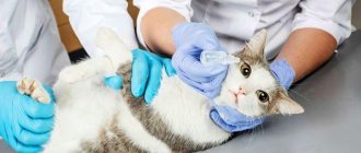
If you see that your cat's eyes are covered with a film, we advise you to contact a veterinarian as soon as possible. Timely treatment will help quickly restore vision and avoid re-infection.
A specialist doctor uses an ophthalmoscope to determine the cause of the underlying pathology of the eye disease and propose an effective course of treatment. In some cases, surgery may be required.
It is imperative to monitor the dynamics of the disease.
Features of intraocular ophthalmomyasis
In some cases, the larvae perforate the tissues of the eye and penetrate under the mucous membrane. In this case, parasitism is called obligate, that is, the larvae must live in such conditions until maturation.
Depending on the specific location of the larvae, anterior and posterior internal ophthalmomyasis are distinguished.
In the first case, the larva is located in the anterior chamber of the eyeball. At the same time, visual acuity decreases and complete blindness can occur. Sometimes the larva penetrates deep into the eye through the sclera, sometimes the routes of penetration are blood vessels or the optic nerve.
With posterior internal ophthalmomyasis, the disease can be asymptomatic, but in some cases it manifests itself as sudden blindness. During examination, the doctor may identify signs of optic neuritis, retinal detachment or other pathologies.
Treatment
To treat this type of disease, veterinarians offer several methods:
- drops or ointments with an anesthetic effect - they help relieve inflammation;
- inclusion of vitamins B12 in the cat’s diet;
- balanced diet;
- washing the eyes using olive oil, warm tea, boric acid or just warm water.
The simplest method, but no less effective, is considered to be preventive eye rinsing. This procedure makes it possible to remove dust and foreign particles from the eyes.
How to wash your eyes
The rinsing procedure is required periodically when the eyes become watery, suppuration appears, or a film forms. Before starting, you should wash your hands thoroughly with soap to avoid further infection. It is better to rinse with an assistant: one person fixes the pet, the second acts, this way the process will go faster.
You need to use a special solution to wash your eyes. Rinsing is carried out as follows: thoroughly moisten a cotton swab in the solution and then carefully rinse both eyes of the animal. After completing the procedure, be sure to blot your eyes with a dry cotton pad.
How to help your beloved cat: prevention
The disease will not develop if you begin to take systematic preventive measures in time. You can help your pet by trying to protect it from the risk of eye damage or infection in this organ by treating it with special solutions.
For these purposes, experts recommend using chamomile decoction - two filter bags infused in two hundred milliliters of water. Use a cotton pad or swab to rinse the animal’s eyes, and then thoroughly wipe the area around the eyes with an additional cotton pad.
Preventive measures also include reviewing the cat’s diet. The menu should contain only high-quality products. Sweets and processed foods are considered off limits for these furry creatures.
It is recommended to visit a specialist to identify pathologies at an early stage and successfully treat them.
Prevention measures
The most effective way of therapy is to prevent the problem.
To prevent inflammation of the third eyelid in a cat, use the following recommendations:
- Create a balanced cat diet from quality products.
- Be sure to add vitamins to your food.
- Periodically carry out eye rinsing procedures.
- Monitor your pet for changes in behavior and examine its eyes regularly.
- Visit a veterinarian at least once a year.
Remember, the sooner you detect eye disease in a cat, the greater the likelihood of its speedy cure. You should not self-medicate; you should immediately take the animal to the veterinarian.
Acute and chronic form
Keep in mind! In adult patients, dacryocystitis can develop in acute or chronic forms.
The two types of disease can be distinguished by symptoms, and in the acute form the following symptoms can be observed in the patient;
- increased body temperature;
- swelling in the area of the lacrimal sacs ;
- narrowing of the palpebral fissure;
- possible swelling of the eyelids ;
- pain in the orbit of the eye;
- manifestation of general symptoms of intoxication of the body.
The tumor , which can be easily felt in the area of the lacrimal sac, may initially be dense to the touch , but after a few days it begins to soften and the swelling subsides.
During this period, an abscess forms, which can spontaneously open, and due to the outflow of pus from it, swelling decreases.
In the chronic form, the patient does not experience pain , but at the same time he experiences strong constant lacrimation , and the swelling in the area of the lacrimal sac turns into a tumor, upon pressing on which pus begins to flow out of the lacrimal canaliculi.