Clinical manifestations of dysplasia Diagnosis Treatment Until recently, it was believed that cats were not susceptible to hip dysplasia (HJD). This disease is widespread among many dog breeds as a hereditary abnormality. According to new information, it turned out that this disease also occurs in cats and is most often inherited. The development of dysplasia in a cat or dog depends not on one gene, but on a whole complex of genetic factors. If dysplasia is detected in a dog or cat, it means that one of the animal’s parents is also a carrier of this disease.
The term "dysplasia" refers to abnormal tissue development. The hip joint consists of convex and concave surfaces. The convex surface is the tip of the femur (head) that fits into a socket formed in the pelvic bones called the acetabulum. In a normally developed joint, the femoral head is completely aligned with the acetabulum, allowing the joint to function smoothly and efficiently. The large muscles of the hip and pelvis help hold the joint in place and allow it to function properly.
With dysplasia, the components of the hip joint are not formed correctly, so the head of the femur does not coincide with the acetabulum. This leads to the head becoming more mobile in relation to the joint. Over time, this abnormal mobility and joint degeneration leads to chronic changes in the hip bones. In most cases, both hip joints are affected, although one may be more affected than the other.
What is joint dysplasia
Each joint is formed by the articular surface of the rounded ends of the bones, surrounded by an articular capsule. Inside there is an articular cavity filled with synovial fluid. Normally, all components of the joint correspond to each other and are firmly held by ligaments.
With dysplasia, due to developmental deficiency, the mobility of bones inside the joint increases, a pathological gap is formed between them, as a result of which the joint cannot fully function.
Description of the disease
Translated from Greek, dysplasia means “disorder, change in shape.” And this fully reflects the essence of the disease, in which improper development and formation of a tissue or organ occurs.
© shutterstock
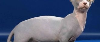
Previously, it was believed that only dogs were susceptible to dysplasia, but over time, this pathology began to be diagnosed in cats, especially purebred ones. According to statistics, large breeds of cats suffer from dysplasia. Thus, among Maine Coon representatives the probability of developing pathology is 18%. Breeds such as Norwegian Forest, British, Scottish, and Persian are also at risk.
Cats are diagnosed with two types of joint dysplasia:
- Hip dysplasia is the most common and affects the lower limbs of the animal.
- Elbow dysplasia is much less common and is a pathology of the forelimbs in cats.
Hip dysplasia in cats can occur at an early age, which has a significant impact on their later life. In this condition, the femur does not align properly with the pelvis. This means that the head of the femur moves relative to the pelvic cavity and a free space appears between them. With this pathology, the connective tissue is the first to be affected, and then the bones are deformed.
With forelimb dysplasia, the bones may not fit together due to their shape or size. In addition, microcracks may appear in the lower part of the bones or they may increase due to salt deposition.
Due to improper development and deformation of the joints, excessive mobility occurs. Over time, the friction of the joint elements increases, and the pressure on them increases significantly. As a result, bone and cartilage tissue are destroyed and many diseases of the musculoskeletal system develop.
Congenital and acquired dysplasia
Cats have congenital and acquired joint dysplasia. The largest percentage of cases is a congenital anomaly that occurs during embryonic development. But at present, scientists cannot identify a specific gene responsible for the development of dysplasia. They believe that this defect is formed under the influence of a specific set of chromosomes or a certain sequence of their connection.
According to another hypothesis, dysplasia is provoked by the synthesis of an insufficient amount of hyaluronic acid, which is part of the synovial fluid. This indicator is laid down at the genetic level, and it is impossible to influence it by external factors.
Acquired dysplasia can be caused by:
- intensive growth of kittens;
- an unbalanced diet, when the body receives an excess amount of phosphorus with a lack of calcium;
- birth injuries.
Note! The most intensive growth in the first months of life is observed in cats of the Asher breed. And although the debate about the recognition of this breed still does not subside, when choosing a pet, owners need to approach the assessment of its constitutional characteristics “with passion”, and also very carefully select the diet.
List of medications for dysplasia
In conservative therapy, medications are used to relieve symptoms and normalize the cat’s condition.
List of medications:
- Stride plus.
- Arthroglycan.
- Akti-Vet.
- Chionat.
- Discus Compositum.
- Hondraton.
- Rimadyl.
- Ketofen.
- Stop arthritis.
- Bonharen.
- Previcox.
- Target.
Additionally, intra-articular injections based on hyaluronic acid are used in treatment. Non-steroidal medications, anti-inflammatory, painkillers (Traumel), symptomatic medications, vitamins, mineral complexes, and alternative medicine may be prescribed.
Important! All medications, course of treatment, dosages should be prescribed only by verinar.
Causes of dysplasia in cats
Owners should understand that genetic predisposition to the disease is not a death sentence. Thanks to proper feeding and maintenance, the degree of development of pathology can be minimized. Therefore, you need to know the reasons for the development of dysplasia in cats in order to avoid mistakes in care.
- Obesity. Excess weight significantly increases the load on the animal's osteoarticular apparatus, leading to its deformation. In addition, an obese pet leads a sedentary lifestyle, as a result of which the muscles and ligaments weaken and no longer fully support the skeleton. Therefore, any sudden movement or physical activity can provoke pathological changes in the joint, leading to dysplasia.
- Injuries. After bone fractures, joint dislocations, sprains (ruptures) of ligaments, muscles or tendons, a natural redistribution of weight occurs, during which the load on healthy limbs increases, and, as a result, the risk of developing joint dysplasia increases. If during an injury the joint capsule was damaged and synovial fluid leaked out, then irreversible changes will certainly occur in the joint, leading to dysplasia.
- Low level of physical activity. Even pets with normal weight should have a well-developed muscular corset that holds the articular surfaces of the bones at a certain distance from each other. With low muscle tone, compression of articular cartilage and menisci occurs, protrusions, hernias and dysplasia occur.
- Early sterilization. The optimal age for sterilization of cats is considered to be from seven months to two years. Removing the gonads too early leads to changes in overall hormonal levels, slower bone growth and the deposition of subcutaneous fat.
- Rickets. Insufficient intake of calcium and vitamin D during the period of intensive growth and intrauterine development of kittens leads to the development of rickets. It manifests itself as curvature of the spine, deformation of the limbs and chest.
Note! Vitamin D is not synthesized in the skin of cats under the influence of sunlight, as it happens in humans. Therefore, it makes no sense to “replenish” your pet’s reserves of this vitamin by walking in the fresh air.
- Hormonal disorders. Impaired bone mineralization occurs when the thyroid gland malfunctions, since it is responsible for the rate of bone tissue regeneration. Dysplasia can also form due to insufficient production of growth hormone synthesized by the pituitary gland.
- Poor nutrition. Even though cats are carnivores, their diet should include grains, vegetables and fruits. Feeding only meat and meat products does not provide the pet with all the necessary microelements and vitamins, and the lack of fiber negatively affects the functioning of the gastrointestinal tract, the absorption of nutrients and the development of beneficial microflora. It is also worth noting that ready-made economy and premium food do not provide animals with the necessary substances for growth and development, which also leads to osteoporosis and dysplasia.
Risk factors
Hip dysplasia in a cat is caused by a number of possible reasons. The most significant factor is considered to be excess body weight. As a result, joint pathology is more often diagnosed in Maine Coons. However, there are other breeds that are more susceptible to dysplasia:
- British;
- Persian;
- exotics;
- Siamese.
The following risk factors are also identified:
Pathology can develop in an animal that eats only dry food.
- pathology of bone tissue;
- complications after suffering traumatic injuries;
- excess high-calorie foods on the menu;
- low physical activity;
- lack of calcium in the body;
- feeding exclusively with ready-made dry complementary foods;
- hormonal imbalance.
Symptoms and first signs of dysplasia in cats
Characteristic symptoms and the first signs of dysplasia in cats begin to appear during a period of intensive growth. Abnormalities of the elbow joints become noticeable only from the age of six months.
The degree of their manifestation depends on the stage of development of the pathology. The first warning signs are:
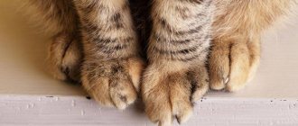
- tension and unnatural gait;
- lameness after sleep, and in severe cases - constantly;
- curvature of limbs;
- muscle atrophy and enlargement of limb joints;
- inactivity;
- a crunch made by joint surfaces rubbing against each other during movement.
Young kittens may meow frequently in pain while moving. They try not to make sudden movements and jumps. Adult animals cannot bend their hind legs under themselves when sitting and hide them under their stomach while lying on a horizontal surface.
Treatment of DTBS
As noted earlier, many cats with TBS do not experience any discomfort. If a cat with this diagnosis is overweight, then regulating its weight will reduce the manifestation of discomfort. For cats with obvious clinical manifestations of THD (lameness, pain), drug treatment is used.
Veterinarians prescribe anti-inflammatory and painkillers, as well as feed additives to restore the joint. Limiting exercise (for example, limiting the animal's going outside or jumping high) will also help treatment. In severe cases, surgical removal of the head and neck of the femur is used, and the damaged tissue of the joint is excised. After this operation, the joint begins to function properly again, and cats experience pain and discomfort as soon as the postoperative recovery period is over.
Physical rehabilitation methods are used as a weight loss measure and a drug-free treatment for hip dysplasia in cats, as in dogs. Rehabilitation is also indicated to restore range of motion and form the muscle frame in the postoperative period.
(c) Veterinary center for the treatment and rehabilitation of animals “Zoostatus”. Varshavskoe highway, 125 building 1. tel.
8 (499) 372-27-37
Breed predispositions
It is worth noting, first of all, such large cats as the Maine Coon. 18% of this breed suffer from dysplasia.
The following are:
- British;
- Scottish fold;
- Siamese;
- Persian;
- Norwegian forest
Life expectancy of cats with OCD
Of course, all owners of Scottish fold cats with osteochondrodysplasia are concerned about the life expectancy of a pet with such a disease. Fortunately, the disease does not directly affect life expectancy. When the first symptoms of OCD appear, owners should take their cat to the doctor as soon as possible. After all, timely treatment can slow down the progression of the disease and improve the pet’s quality of life.
Some veterinarians suggest that owners euthanize a sick pet or amputate the affected limb. However, with good care, supportive care and patience on the part of the owner, the condition of the Scottish Fold cat can be significantly improved. You can also increase your pet's lifespan with a balanced diet.
Unfortunately, if the disease is too severe, it is still better for the animal to undergo euthanasia. It is suggested to do it if the disease is constantly progressing, the pet does not get up at all and does not go to the litter box, and he also has to take pills that no longer bring relief. In such a situation, it is better to stop the cat’s suffering, but the decision always remains with the owner.
Tests and methods for diagnosing dysplasia
It is impossible to make an accurate diagnosis and begin proper treatment using clinical signs alone at home. Therefore, you should take the animal to the clinic, where tests will be prescribed.
- Collection of anamnestic data. During the survey, the specialist examines the pedigree (if any), finds out the presence of the disease in the parents and other kittens from the litter, the characteristics of pregnancy and childbirth, the presence of birth injuries, and the time of the first signs.
- Clinical examination. It includes examination of the limbs and spinal column, palpation, determination of the presence and degree of bone deformation.
- Laboratory analysis of blood and urine. An increased number of leukocytes indicates the development of inflammation in the body and the presence of associated pathologies.
- Study of the functioning of the cardiovascular system. Since general anesthesia is required for radiography, before administering it the doctor must make sure that the animal does not suffer from heart disease.
- Arthroscopy. During this procedure, a specialist can assess the condition of the inner surface of the joint using an arthroscope inserted into it through a pinhole puncture.
Note! To conduct this examination, you need expensive equipment, appropriate qualifications and experience of specialists. Therefore, not every clinic can afford such a “luxury”. In addition, the high cost of the study does not make it possible to make it widespread and publicly available.
The final diagnosis is made based on radiography of the joint.
Symptoms
Age-related changes appear in growing pets. For example, elbow deformity develops within six months and is immediately accompanied by symptoms.
Signs of the disease depend on the severity of the pathology:
- the appearance of an unsteady gait;
- the cat stops jumping on high surfaces;
- kittens develop lameness (at first - when the pet lies in one position for a long time, as the pathology progresses - the symptom becomes permanent);
- the paws become bent and become X-shaped;
- a frisky cat stops moving quickly;
- muscles begin to atrophy, accompanied by weakness of the paws and weight loss of the pet;
- thickening at the bends of the limbs;
- the cat cannot sit in the “Egyptian” pose;
- when the pet lies on its stomach, spreads its paws to the sides;
- A crunching sound can be heard when the cat moves.
If the initial stage of the disease is not treated and osteoarthritis begins, the pet begins to move with great difficulty or completely loses the ability to walk independently.
Test for dysplasia in cats
In 1983, scientists proposed a test for dysplasia called PennHIP. After completion in 1993, it became widespread, as it allows, with certain equipment, to recognize pathology at the earliest stages of development.
After general anesthesia and muscle relaxants that relax the skeletal muscles are administered, the cat is fixed on a special device in a stretched state. This makes it possible to move the femoral head away from the acetabulum to the maximum distance.
The doctor then takes several x-rays and calculates the DI displacement index by comparing the data obtained with the breed standard.
- A DI value of “0” indicates a severe degree of dysplasia and the presence of degenerative processes.
- DI=1 indicates low joint mobility.
- With an average degree of pathology, this coefficient is 0.5-0.6.
X-ray of the joint
The main method for diagnosing dysplasia today remains x-ray of the joint. It allows you to accurately assess the degree of tissue damage and bone condition.
The disadvantages of this survey are:
- age restrictions (the pet must be 2 years old at the time of the procedure);
- the need to administer general anesthesia to ensure complete immobility.
Note! If a cat suffers from heart pathologies and at the same time has a calm disposition, then radiography can be performed without anesthesia, but with careful fixation.
Juvenile symphysiodesis
The operation is quite simple and low-traumatic. The essence of the procedure is that it is necessary to partially block the growth zone along the symphysis of the pubic bones. Using an electrocoagulator, the symphysis is cauterized at intervals of 2-3 mm, power 40 W, exposure time - 10 seconds at each point, using an electrode in the form of a spatula. If a needle-shaped electrode is used, the exposure is 30 seconds. Since the procedure is performed at a young age, during the period of most active growth, the acetabulum spontaneously unfolds around the femoral heads due to slow growth in the symphysis area.
Rice. 5. Juvenile symphysiodesis.
The best results are observed in those animals who underwent this procedure at 16 weeks. The method is ineffective after 20 weeks.
The main advantages of the operation are its low-traumatic nature, low cost and complete restoration of weight-bearing ability. The main disadvantage is the very short period in which this procedure can be carried out. It is difficult to convince the owner of an animal who does not yet have any clinical problems to decide on this operation. Due to the lack of awareness among dog owners (few people check their pet for the presence of dysplasia at 3-4 months), the procedure is rarely performed, so we have to deal with really sick patients who already need surgical help.
Consequences of the disease
Conservative methods cannot completely restore the integrity of the joint and ensure its full functioning. Therefore, the consequences of the disease are:
- joint deformation;
- degeneration of bone and cartilage tissue;
- secondary osteoarthritis;
- hip subluxation;
- hip dislocation;
- pelvic deformity;
- arthritis;
- complete loss of joint mobility.
As a result of these complications, cats become very difficult to move and experience increased pain.
Differential diagnosis
Differential diagnosis was carried out using the classic DAMNITV mnemonic method (Appendix 1). The initial suspicion of trauma (history, absence of tail, dirt, impaired movement) was rejected due to radiographic examination.
Appendix 1. Mnemonic method used in differential diagnosis called “DAMNITV”
D - degeneration A - anomaly (congenital) M - metabolism N - neoplasia (nutritional) I - inflammation/infection/ideopathic/iatrogenic nature T - trauma/toxicosis V - vascularization
The complete absence of pain reaction made it possible to exclude injury in the area of the lumbosacral joint of the spinal column, as well as spondylodiscitis and inflammation of the spinal cord. It is unlikely that the animal could have a neoplasm at this age. The assumption, based on the results of a neurological study (paresis, jumping, ataxia, etc.), was focused on an anomaly of a congenital nature. The absence of a tail, as well as the results of a clinical and complete neurological examination with the addition of an x-ray, led to the conclusion that we are talking about dysplasia at the level of the sacro-caudal joint. This congenital anomaly is often found in individuals with a short tail or no tail, such as the Manx breed.
Prevention measures
The main measures to prevent dysplasia are:
- culling and sterilization of kittens with a genetic predisposition to the disease;
- feeding pets with high-quality food, balanced in the content of nutrients, vitamins, calcium, phosphorus and other minerals;
- preventing obesity;
- regular examination by an orthopedist.
The cat should have a soft bed. It should be placed in a warm place where there are no drafts, which can provoke inflammatory processes in the joints.
Causes
Osteochondrodysplasia in cats is inherited. Its manifestation is not related to the gender of the animal.
OCD is considered a disease of Scottish Fold cats, because they are the owners of a mutant gene for fold ears. To avoid the appearance of offspring with osteochondrodysplasia, breeders always cross fold-eared cats with straight-eared cats. In this case, the risk of a dangerous gene appearing is minimal. However, in rare cases, even crossing a straight with a fold may result in a kitten with osteochondrodysplasia. That is why in some countries it is prohibited to breed Scottish Fold cats.
In Scottish Folds, the disease varies in severity. The most common disorders are the development of the bone skeleton, but in the most severe situations, deformation of the limbs can develop.