One of the reasons why people love pets is their warm, soft fur that is pleasant to stroke. In addition, the coat is a source of pride for many types of cats and dogs, making them beautiful and easily recognizable. However, there are often cases when, due to various ailments, the wool loses its quality, becomes excessively greasy, falls out, or becomes tangled. Veterinarians note that one of the common reasons why feline lovers come to them is a cat’s greasy tail. In this article we will talk about why a cat gets the greasy tail effect and how to deal with it.
Causes of the disease
Cats are graceful and graceful animals, which are very neat and well-groomed. Therefore, it is doubly offensive when a pet has oily hair on its tail. This spoils the impression of your four-legged pet. There are several reasons for the appearance of such a defect - internal and external; let’s look at each of them in more detail.
Domestic
You need to start with the reasons that arise due to malfunctions in the cat’s body. Fortunately there are not so many of them:
- Hyperfunction of the paraanal glands. They are located at the very base of the cat's tail, their function is to lubricate the animal's anus, which makes it easier for him to defecate. When the outlet becomes inflamed or narrowed, there is a delay and accumulation of a foul-smelling substance in the anus. It is important to know that pets that have not been castrated are more likely to develop this disease. Experts say that a possible reason for this is the pet’s elevated hormone levels.
- Dysfunction of the sebaceous glands, which are located directly on the cat’s tail. If their functioning occurs too intensively, then the fat does not have time to spread evenly throughout the pet’s fur, concentrating in one place. In a neglected state, this leads to complete blockage of the glands, which creates a favorable atmosphere for the development of harmful microorganisms. Veterinarians notice that active production of keratin often leads to this. At the site of the lesion, a characteristic black spot may appear - a comedon. Over time, if an infection gets there, it threatens to develop into an abscess on the cat’s skin.
- Hairless and short-haired breeds of cats fall into a special risk group, which is characterized by the effect of a fat tail. It is quite difficult for them to identify it.
External
In addition to internal reasons, there are also external ones that can contribute to the formation of excess fat on a cat’s tail. These include:
- Poor animal hygiene. If a pet is overweight, this makes it difficult for him to attempt self-care. An aggravating factor can be the owner’s inattention to such issues, as well as the cat’s categorical dislike of taking baths.
- Poor nutrition, lack of vitamins. These two factors seriously affect the level of subcutaneous fat production. Therefore, the owner of a “problem” cat needs to carefully monitor his pet’s diet.
In any case, only an appropriate specialist can give a qualified assessment of the reason why a cat’s fur becomes greasy. Do not try to diagnose yourself, this will aggravate the situation, which will make the animal suffer and become even more unsightly.
Ulcers
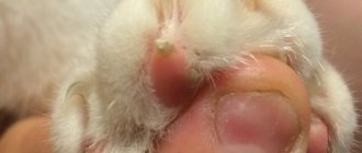
More often, ulcers occur in short-haired four-legged friends. They can appear along the entire length of the tail, often affecting the root, and this can cause necrosis of the tissues and vertebrae of the tail.
An ulcer on the tail requires serious treatment and attention from the owner. If a crust has formed on it, then it must be removed from there and the wound must be cleaned of suppuration. Be sure to treat everything with an antiseptic for cats. Also, it is worth treating it with ointment (Vishnevsky, streptomycin) and applying a bandage.
© shutterstock
In advanced cases, the cat may develop gangrene, blood poisoning and serious consequences.
Symptoms of the disease
Fat tails are much more common in male cats than in male cats. As noted above, animals with short fur are at risk, which complicates the diagnostic process. Let's try to find out what signs clearly demonstrate this disease:
- The epithelium at the base of the fur becomes lumpy, it flakes off and becomes covered with a thin crust of pus.
- The tail, most often at the junction with the body, is thickly covered with fat, which makes the fur begin to shine. The discharge cannot be washed off with shampoo or soap.
- If the disease progresses, the skin at the site of localization will be combed and covered with red pimples. This is how the animal tries to relieve the itching, but only spreads the infection through the skin.
- The area of “greasy” fur becomes exposed over time. This is due to the fact that the hairs gradually break off at their base.
- A symptom that the anus is inflamed may be uncharacteristic behavior when the cat crawls with its butt on the carpet, trying to release excess contents.
- Over time, brown spots may appear on your pet's tail; if left untreated, they will develop into a hard crust, tearing which the cat will injure itself.
Owners who notice that their animal has developed “greasy tail” syndrome should under no circumstances dismiss this problem, attributing it to seasonality or simply the cat’s untidiness. Most often, the true cause lies precisely in a serious illness that has just begun to manifest itself. If you react promptly, it will save a lot of hassle for both the owners and the cat.
Prevention
- Sterilization helps to avoid problems with the greasy tail by ensuring a calm hormonal balance. But it is worth noting that many uncastrated cats never encounter such a dermatological problem.
- A balanced diet and timely consultation with a specialist at the first signs of the disease will help prevent hyperplasia of the sebaceous glands. Preventing obesity throughout a pet’s life and caring for its fur is something any owner can do.
Treatment options
Treating a cat's fatty tail is a long and tedious process. You need to start with a trip to the veterinarian, who will determine exactly what caused the disease and how to eliminate it as quickly as possible. Treatment, as a rule, is based on the fact that the pet begins to take baths more often. To do this, you need to buy special shampoos containing lactic acid. This will perfectly exfoliate harmful keratin accumulations and cleanse the skin, which will create the basis for subsequent therapy.
If the veterinarian determines that the greasy tail is caused by a hormonal disorder, then castration of the pet or medication to stop the production of sex hormones will be required. Excessive production of anal secretions is treated with rinsing. The specialist will clearly explain to the owners how this is done. If the disease recurs regularly, the anal glands can be completely removed surgically.
If the causes are caused by external irritants, then a complex of vitamins and a special diet are prescribed to strengthen the immune system. All this will help the cat become beautiful again, and its owner - calmer. If you ignore the fact that your pet’s tail becomes fat, then in the future this can lead to complete baldness, which will make the animal completely unsightly.
What can trigger seborrhea?
The basis of treatment is determining the cause of the pathology. Seborrhea can be provoked by both local and systemic disorders that provoke general disorders:
- skin diseases - affect the epidermis, which dies and flakes off in the form of dandruff;
- allergy – against the background of immune hyperreactivity, pathological skin lesions are noted;
- parasites – can indirectly provoke the disease;
- systemic pathologies – connective tissue diseases, arthritis, arthrosis;
- endocrine diseases - hyperreactivity of the glands contributes to damage to the epidermis;
- Metabolic disorders – when metabolic disorders occur, complex mechanisms are triggered with skin damage.
In the presence of an underlying disease, signs of a specific pathology appear: for example, parasites contribute to exhaustion against the background of increased appetite; with skin pathologies, rashes, papules or blisters appear.
Recommendations
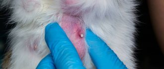
If an abscess is found in a cat, how to treat it at home? It is recommended to show the animal to a veterinarian to make an accurate diagnosis and prescribe a course of therapy. A disease such as an abscess is common in cats. Since it needs to be opened, it is better to do this at a veterinary clinic.
This procedure can be done in two ways: under local anesthesia or general anesthesia. The veterinarian will determine how this operation will proceed after an examination. And depending on the condition of the animal, he will prescribe which painkiller is best for making the incision.
Although abscess is a common condition in cats, hairless cats are less susceptible to it. The reason for this state of affairs is that cats without fur are kept by their owners in favorable conditions. Therefore, they do not have the opportunity to sort things out with other animals and enter areas other than their own.
As a rule, to eliminate the inflammatory process, cats are prescribed antimicrobial agents. These include sulfonamide drugs. It is necessary to give your pet a course of medications. You should not stop taking medications, even if your cat’s abscess appears to have become smaller. Antibiotics should be taken as a course. It is 5 or 7 days. The bottom line is that when the course of drugs is interrupted, the body can develop immunity to their components. And if you need to take these medications again, they will not have the desired effect on the body.
Symptoms of hair loss in cats
Photo.
Baldness of a cat Symptoms of baldness will be noticed even by an inexperienced owner. The cat leaves hair everywhere, some parts of the body become bald (the ears, base of the tail, stomach and back are most often affected).
The condition of the skin determines what exactly caused the development of alopecia. In some cases, the cat develops redness in areas of baldness, the skin itches and worries the animal. In case of serious diseases, scabs and ulcers form, the skin peels off, and sometimes small bumps grow.
Areas of baldness can cause discomfort for your cat. If an animal scratches its skin furiously or does not allow itself to be touched, this is a reason to take it to the veterinarian. In the affected areas, the skin may become hot and stiff.
Why does necrosis occur?
The pathology is characterized by irreversible tissue death and cessation of cell activity due to impaired blood circulation, nerve conduction, and the activity of anaerobic or pyogenic bacteria. Most often, areas of the body where the blood vessels are small are affected - ears, paws, muzzle, tail, tongue. Often internal organs are affected - intestines, stomach, kidneys, liver. Depending on the location, the causes of necrosis are as follows:
- Internal organs: entry of foreign objects into the gastrointestinal tract;
- helminthiases;
- cirrhosis;
- volvulus.
- frostbite;
- glossitis (inflammation of the tongue);
Allergies or exposure to toxic chemicals can lead to tissue death.
In addition, the following factors cause tissue death in any area:
- autoimmune failure;
- allergy;
- prolonged inhalation of pesticides by animals;
- heart attack;
- electrical injury;
- disturbance of nerve conduction.
Abbreviations: APTI—fine needle aspiration; IM – intramuscular; s/c – subcutaneously; p/o – inside; GCS - glucocorticosteroids
Erosions and ulcers can be a manifestation of various skin diseases in cats. Erosion (erosio) is a superficial defect within the epidermis (epithelium), can be found on the skin and/or mucous membranes. Appear due to mechanical irritation of the skin - as a result of scratching (excoriation) of a papular rash, during maceration of the epidermis of the skin in the area of folds (intertrigo complex). Erosion also forms when vesicles, blisters and superficial pustules open. Many immune-mediated diseases accompanied by vesiculobullous lesions go unnoticed by owners until they erode. They are usually found in places with sparse hair (belly, temporal areas). Erosion usually has a bright red color, often covered with discharge or crusts. A distinctive feature of erosions is healing without scar formation. Sometimes temporary pigmentation appears in their place, less often - hypopigmentation. Erosion causes itching and sometimes even pain. Cats lick the affected areas intensively; maceration and infection lead to the development of ulcers.
An ulcer (ulcus) is a deep defect of the skin, and sometimes of the subcutaneous tissue. Acute ulcers are usually shallow, usually round or oval in shape, their edges do not rise above the level of the skin or mucous membrane. The edges of a chronic ulcer are often raised, dense, and sometimes calloused (callous ulcer). The bottom, or base, of the ulcer can be clean, bleeding or suppurating, covered or not covered with granulations. In a number of diseases, it is covered with necrotic decaying masses. With pronounced cicatricial changes in the area of the edges and bottom, the ulcer often acquires a peculiar star-shaped outline. Subsequently, a star-shaped scar can be found in their place. Skin ulcers are a polyetiological disease; they can occur as a result of the development of infection (bacterial, viral, fungal), mechanical, thermal, electrical, chemical factors, and ulceration of tumors.
Diagnosis of erosive-ulcerative dermatoses is carried out on the basis of a medical history, a dermatological examination, including the study of deep scrapings from the skin, and routine blood tests. One should not neglect the collection of anamnesis when examining cats; for example, the development of skin ulcers can be caused by direct damaging effects due to thermal damage, as a result of exposure to acids, caustic alkalis, etc.
Cytological examination of scrapings and material obtained by fine needle aspiration (FNA) should be performed at the initial stage of the diagnostic examination (before applying therapy). This allows you to get a quick result and early diagnose cutaneous neoplasia, although the absence of tumor cells in the material does not exclude its presence.
It is necessary to carefully select for study the most “fresh” affected areas (usually at least three) that are not subject to excoriation (scratching). In ulcerative dermatitis, it is preferable to find primary lesions, which are a direct result of the pathological process and provide the most information. Before taking scrapings, purulent-necrotic masses must be removed from the surface of the lesions with a cotton swab (gauze pad). The material is scraped off using a scalpel blade until capillary blood appears. Next, it is evenly distributed over a glass slide and dried in air.
Methods for processing and painting smears, punctures and prints are very diverse (according to Pappenheim, azure-eosin according to Romanovsky, according to Leishman, hematoxylin-eosin, fast Diff-Quick paints and others).
In hematological blood tests of cats with erosive and ulcerative lesions, attention is paid, first of all, to the number of leukocytes and leukogram: leukopenia is observed in viral diseases (calicivirus, feline immunodeficiency, etc.), eosinophilic leukocytosis - with increased sensitivity to flea bites, atopy, eosinophilic granuloma, especially with the presence of ulcers in the oral cavity, less often food allergies, urticaria pigmentosa and mastocytomas.
Biochemical blood tests are often used to diagnose systemic disorders to exclude diabetes mellitus, renal and liver failure. Identifying these metabolic disorders limits the doctor's choice of medications, which helps avoid complications when treating sick cats. When diagnosing skin diseases of an erosive-ulcerative nature, attention is paid to the proteinogram, especially the content of immunoglobulins - pronounced hypergammaglobulinemia is observed in long-term allergic diseases, for example, atopy.
Diseases of an immune nature
Eosinophilic granuloma complex is the most common erosive and ulcerative skin disease of cats, affecting the skin and oral cavity (photo 1). Clinically manifested by ulcers, plaques, linear granulomas, miliary dermatitis (photo 4). In cytological preparations, a large number of eosinophils and histiocytes are observed (photo 3). To obtain informative material, it is necessary to carry out scrapings from the most recent areas of the lesion. In cases of infected erosions, examination of the material reveals degenerative neutrophils, microbes, and cellular detritus. Imprint smears of superficial exudate almost always contain many bacteria and leukocytes, which indicates the colonization of opportunistic microorganisms in the ulcerated lesion.
In plasmacytic pododermatitis, cytology of aspirates obtained from eroded granulomas located mainly on the paw pads reveals a large number of plasma cells.
Mixed inflammatory cells represented by non-degenerative neutrophils and macrophages, the presence of a large number of acantholytic cells (rounded nuclear keratinocytes, devoid of cytoplasmic processes) are characteristic of an autoimmune process (for example, pemphigus foliaceus - photo 6).
At the beginning of treatment, it is necessary to use mechanical protective equipment to prevent self-injury. This could be an Elizabethan collar, light overalls, soft polymer caps for claws and other devices.
GCS and immunosuppressants are used to treat immune-mediated erosive-ulcerative dermatoses in cats. Corticosteroids have a rapid anti-inflammatory, anti-edematous, antipruritic effect, inhibit the release of cytokines (interleukins and interferon) from lymphocytes and macrophages, inhibit the release of inflammatory mediators by eosinophils, disrupting the metabolism of arachidonic acid and the synthesis of prostaglandins, reduce inflammatory cellular infiltrates, reducing the migration of leukocytes, including . lymphocytes to the area of inflammation.
Prednisolone is prescribed po 1 - 2 mg/kg every 12-24 hours, this interval gradually increases to 48 - 72 hours. It is important to continue treatment for at least a week after visible remission has been established. If it is impossible to give tablets, you can use prolonged injectable drugs GCS: dexamethasone 1 - 2 mg (Dexafort, MSD Animal Health, subcutaneously, intramuscularly at a dose of 0.3 - 0.7 ml), methylprednisolone acetate (Depo-Medrol, PFIZER ) is administered intramuscularly in a dose of 0.25 – 0.5 ml (10 – 20 mg per cat), triamcinolone (Kenalog, Polcortolone 40 – intramuscular dose 0.2 – 0.5 ml per cat). After the administration of GCS, in most cases, improvements are observed within 12 to 48 hours, and the effect lasts for several days or even weeks. However, due to the risk of developing diabetes mellitus and immunosuppression, it is advisable to avoid frequent use of prolonged injectable forms. Antihistamines and fatty acid supplements may be used in addition to steroid medications. For humans, antihistamines in monotherapy, including local ones, are considered effective, but, in the author’s experience, they are of limited value for cats, although in some cases they reduce the need for GCS.
If it is impossible to exclude exposure to the allergen and the disease recurs when the dose and frequency of administration of GCS is reduced, combination therapy using cytostatics is used.
Drugs in this group are used to treat cats with pemphigus and eosinophilic granulomas that are not amenable to GCS therapy. More often, chlorambucil (Leukeran) is used in doses of 0.1-0.2 mg/kg (2-4 mg/m2 body surface) daily or every other day. Symptoms of gastrointestinal tract damage (anorexia, vomiting, diarrhea) occur less frequently when prescribed every other day. Myelosuppression disappears after discontinuation of the drug. The course is usually 1–2 months, then the dose can be reduced until the drug is discontinued. When treating animals with drugs of this group, it is recommended to monitor the clinical blood test, including platelet count, every 2 weeks.
For eosinophilic dermatitis, cyclosporine is successfully used at a dose of 2.5 mg/kg/day. In severe cases, the dose may be increased to 5 mg/kg/day. When a positive clinical result is achieved, the dose should be gradually reduced until completely discontinued.