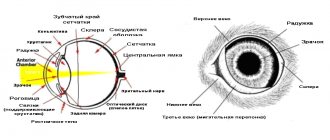
Cats' eyes are very sensitive. Thanks to this, they have a unique feature - to see in the dark. Due to the special structure of the retina, the cat’s pupil reacts sharply to lighting - it expands in the dark, almost covering the iris, or narrows to a thin strip, preventing light damage to the eyes.
But there are situations when the pupil remains dilated, regardless of the lighting. What causes dilated pupils in a cat? Let's try to understand this situation.
The normal state of a cat's pupils
A cat's pupils are always dilated at night, regardless of their internal state. This reaction helps the animal see better in the dark. Surprisingly, cats can even catch the light of stars.
One of the reasons for dilated pupils in a cat is physiology, or rather estrus. If during this period the animal’s pupils are constantly dilated, then this is the norm. This reaction of the body manifests itself to the hormonal changes that occur in the body during this period. At this time, dilation of the pupils is accompanied by signs of the sexual cycle - loud meowing, pressing to the floor with the tail raised up, frequent urination and constant licking.
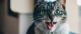
Stress, overexcitation, fear, and aggression are also the causes of dilated pupils in a cat for a long time, regardless of lighting conditions. Experts say that cats see everything in slightly blurred outlines, and when the animal is alarmed or frightened, it intuitively looks around, trying to see sources of danger.
The reason for the cat's dilated pupils in this situation is adrenaline, which is produced whenever the animal is in an excited state. In addition to dilated pupils, such nervousness may be accompanied by hissing and flattening of the ears. Enough for the cat to calm down, the pupils “relax” and again become dependent on light.
Perhaps the owners know another reason for dilated pupils in a cat. When running, active games, jumping, trying to catch some living creature, the cat’s pupils are dilated. The animal needs to clearly see its potential prey, and also measure the distances between objects. This is a completely normal reaction to the production of active adrenaline. After the end of the games, the condition of the pupils will return to normal.
In what cases should you not worry?
The causes of photophobia in cats are not always accompanied by pathology. Newborn kittens suffer from physiological photophobia, as their eyes continue to develop after birth.
After the first attempts to open their eyes, babies may turn away from bright light sources, trying to hide in the far and dark corner of the box. This type of photophobia is not considered by veterinarians and professional breeders as a pathology.
Another natural cause of photophobia, which should not cause concern to the owner of the animal, is the period of pregnancy by the cat. Some animals, in anticipation of replenishment, become affectionate and sociable, while others, on the contrary, try to hide away, hiding their irritation.
This behavior is determined by a natural predisposition, since in the wild cats make their nest in dark places.
Another physiologically justified cause of photophobia in a cat can be a stressful situation. Fear and emotional shock lead to the pet’s desire to retreat to a dark place, avoiding direct rays.
The most common causes of stress in an animal are:
- moving from permanent place of residence;
- long trips;
- going to the hospital;
- the arrival of a new pet in the house;
- lack of attention from the owner;
Such conditions, if properly organized, do not require the intervention of a veterinary specialist and do not lead to serious disorders in the pet’s body. You should carefully consider the reasons for the behavior and try to neutralize them.
Dilated pupils in a cat after surgery
This condition should not alarm owners. Indeed, the dilated pupils of a cat after anesthesia do not react to light for some time. Within a day, their condition returns to normal. Veterinarians recommend leaving the animal in the clinic to monitor recovery from anesthesia for two to three hours. In severe cases, the length of stay in the clinic is determined by the veterinarian.
After castration, the cat's pupils are dilated for 24 hours. After anesthesia, the animal may experience vomiting and severe weakness. You can pick him up from the clinic in an hour.
Eye diseases
If a cat, at rest and in normal lighting, has one or both pupils dilated, then this indicates some kind of pathology in the animal’s health, which should alert the owner. Be sure to take your pet to the vet, as there can be many reasons for this condition. Only a specialist can identify them and make a diagnosis.
It is necessary to understand that the fact of dilated pupils in an animal is not as terrible as the lack of reaction to light.
Why don't my cat's pupils react to light?
It is generally accepted that cats' main senses are smell and hearing, but healthy eyes are an equally important factor for cat health. It happens that owners notice extremely dilated pupils in their pet, but when the light is turned on, no reaction occurs.
Dilated pupils may indicate a problem.
The following two tabs change content below.
- About the expert:
Irina Vladimirovna Shorokhova
I am a veterinarian in one of the clinics in the city of Gomel (Belarus). I myself am an experienced cat lover, I have two Don Sphynxes. I love these animals very much and they love me back. These are charming cats - Marfa and Petrovna.
There may be many reasons for this, but the most dangerous may be glaucoma, retinal detachment, and hypertensive retinopathy.
Glaucoma
A disease characterized by increased intraocular pressure and an increase in the size of the eyeball . This occurs due to the pressure of fluid on the optic nerves, as a result of which these nerves are damaged, which provokes possible blindness. Basically, the pathology is of secondary origin, primary exposure is a rare case.
Glaucoma occurs when intraocular pressure increases.
Symptoms
The symptoms of glaucoma in the early stages are practically invisible, which significantly complicates subsequent treatment.
Next, clouding appears in both organs of vision. If the lesion is unilateral, a noticeable difference in the size of the eyes gradually becomes noticeable. Strabismus develops, the pupil dilates, stops responding to light, and blindness gradually progresses. As the stage progresses, swelling and protrusion occurs.
Glaucoma causes clouding of the eye.
Treatment
It is impossible to completely cure glaucoma, but you can provide your pet with a fairly comfortable existence.
Pilocarpine is dripped, dorzolamide and thymol . It is also possible to prescribe steroids, diuretics, vitamins, and painkillers.
Pilocarpine drops are used for treatment.
Retinal detachment
The mechanism of the pathology is the separation of the retina from the fundus of the eye.
This disrupts the blood supply, which interferes with the normal functioning of the retina. One of the reasons is chorioretinitis, a disease caused by infectious peritonitis, leukemia, and arterial hypertension.
Impaired blood supply provokes retinal detachment.
Signs
Clinical signs are not particularly specific, so the basis of diagnosis is differentiation. Main symptoms:
- partial or complete loss of vision;
- lack of reaction to light;
- the presence of floating fragments of the retina and blood vessels;
- accumulation of blood streaks inside the vitreous;
- dilated pupil.
Retinal detachment causes vision loss.
In addition to the symptoms mentioned, it is very important to distinguish the accompanying signs of the disease that caused the detachment.
Treatment
Retinal detachment is characterized by the most acute course due to the fact that the animal goes blind extremely quickly.
Therefore, the effectiveness of the therapy used will depend on the speed of the host’s response.
Drugs that reduce systemic increases in blood pressure, antifungals, glucocorticoids, and diuretics . In most cases, the only remedy is retinopexy.
The surgical method is used for treatment.
Hypertensive retinopathy
A disease that is characterized by damage to the retina and choroid due to chronic processes in the kidneys or hyperthyroidism.
Provoking factors for the development of primary ailments are violations of the conditions of keeping pets - ignoring vaccinations, routine medical examinations, unsanitary conditions.
Cats living in unsanitary conditions are at risk.
Signs
Signs of hypertensive type retinopathy are expressed in a gradual decrease in vision, which subsequently leads to complete blindness.
The pupils are maximally dilated and there is no reaction to light. Bloody veins are visible inside the eyes. Blood pressure is increased, weakness, apathy, and loss of coordination of movements occur.
The cat becomes weak and apathetic.
Symptoms and treatment
Symptoms of primary origin may also be present - nausea, vomiting, difficulty urinating, refusal to feed due to severe thirst .
There may be a urge to vomit.
Therapeutic measures are based on eliminating the primary factor. At the same time, symptomatic treatment is carried out - painkillers, elimination of fluid deficiency. Removal of intoxication, ophthalmic local remedies.
Anisocoria
With this disease, the dilated pupils of the cat do not react to light. This is a serious reason to visit a veterinary ophthalmologist. The causes of the disease can be not only in the eyes themselves, but also in problems with the nervous system, in particular pathologies of the optic nerve or brain. This condition can lead to blindness if the cause is not eliminated in a timely manner (if the pathology is curable).
The causes of the disease include:
- posterior uveitis;
- retinal atrophy;
- lens luxation;
- glaucoma (angle-closure);
- brain tumors (rare);
- cerebrovascular accident;
- optic nerve atrophy;
- increase in intracranial pressure;
- disruption of the blood supply to the eye.
Photophobia
Photophobia or fear of light is a condition of increased sensitivity of the eye to light. Photophobia causes pain and discomfort in the animal, causing it to squint its eyes and flee to dark rooms or corners.
As a rule, blepharospasm and photophobia are caused by an infectious disease of the respiratory tract or ailments of the eye itself.
Photophobia in a cat can be caused by mechanical injuries or chemical and thermal burns. Also in veterinary medicine, photophobia is often diagnosed due to a foreign object entering the eye or an allergic reaction. Photophobia may develop against the background of progression of tumors in the brain, cerebral edema and neuralgia.
It is necessary to contact a veterinarian for an accurate diagnosis and appropriate treatment.
Consequences of injuries
When scarring of injuries occurs, opacities of varying intensity appear on the cornea. Because of this, insufficient light intensity reaches the retina. Behind the damaged cornea, the pupils reflexively dilate further to capture more light for improved vision. Although there is a reaction to light, it is very weak, and it seems that the pupils are constantly dilated.
Cataract
A disease in which there is complete or partial clouding of the lens of the eye. As a rule, this occurs in the elderly animal due to vitamin deficiency, diabetes or injury. The main symptom is a gray-blue color of the pupil. Treatment is carried out only surgically, but in the early stages and for prevention they use Gamavit and Fitomin.
Blepharitis
Due to external influences or vitamin deficiency, inflammation of the eyelids develops. Consultation with a specialist is necessary to determine the type of disease. Blepharitis can be simple, meibomian, which is characterized by inflammation of the sebaceous glands on the eyelids, ulcerative. The main symptoms of the disease include: thickening of the eyelid, scratching around the eyes, crusts of pus on the surface, dilation of the pupil, and the cat suffers from severe itching.
First of all, the animal’s eyelids are freed from crusts using a solution of soda and oil. Vaseline oil is most effective for softening. The eyelids are treated with brilliant green and mercury yellow ointment. As an auxiliary therapy, the daily diet is enriched with Phytomins.
Conjunctivitis
This inflammation of the eyes is divided into subtypes: allergic, arising due to the ingress of a foreign body, infectious. It manifests itself as liquid discharge from the eyes, often purulent, general restlessness of the animal, photophobia, and the formation of crusts after sleep.
The pupils can both become very narrow and pathologically dilate. With such symptoms, it is very important to provide timely assistance to your pet, independently, but under the supervision of a veterinarian. The cat's eyes are washed with a weak solution of potassium permanganate or Miramistin. Then drops of “Iris” are instilled. If crusts appear around the eyes, they should be soaked and removed with a damp cotton swab. Then the lacrimal canal is washed generously with drops of “Iris” or “Neoconjunctivet”. The procedure is repeated three times a day until recovery. The course of treatment lasts ten days.
For conjunctivitis caused by infection, your veterinarian will prescribe antibiotics. As an addition to the treatment of moderate and mild forms of the disease, you can use hygienic lotions from the “Phytoelita” series, as well as medicinal herbal infusions, to wash your eyes.
Diagnosis of possible diseases
The most modern method for diagnosing pathological changes in the visual organs is electroretinography. A method that allows you to accurately determine how your pet sees and how the retina of the eye reacts. Accurate diagnosis is an important part of the subsequent prescription of adequate treatment.
The animal must first be prepared before the study. It is recommended to keep your pet on a fasting diet for at least 8 hours. This is necessary for a blood test. On the scheduled day of your visit to the veterinarian, it is not recommended to use various eye drops for your pet. This can lead to incorrect diagnostic data.
The total time of clinical examination and diagnostic measures takes no more than 60 minutes, including consultation with a veterinary specialist. To make an accurate diagnosis, the following instrumental and laboratory studies are necessary:
- biomicroscopy;
- measurement of intraocular pressure;
- study of the patency of the lacrimal canal;
- Schirmer test;
- ultrasound diagnostics and examination of the retina with capture of the optic nerve.
Based on the data obtained, the specialist develops treatment tactics based on the individual characteristics of the patient.
