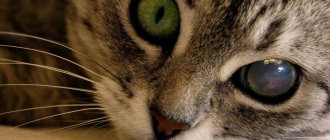
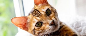
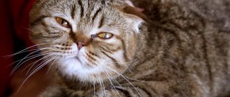
In a healthy cat, the third eyelid becomes visible only when she blinks or tilts her head. The appearance of a light film on one or both eyes that covers almost half of the eyeball is a reason to contact a veterinarian.
Let's find out what functions the third eyelid performs and what pathologies of this organ may arise.
What is the third eyelid
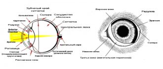
The third eyelid in a cat is a fold of the mucous membrane of the eyelid, which has the shape of a crescent and is located at the inner corner of the eye. Besides the above-mentioned common name, the third eyelid is also called the nictitating membrane or inner eyelid.
A kind of frame for the nictitating membrane is a small elastic cartilage, which is hidden in its folds and has a T-shape, with its “cap” curved in the shape of the eyeball. The lacrimal glands are attached to the “trunk” of the cartilage. They produce most of the volume of tears. Lymphoid tissue is located under the mucous membrane that covers the inner eyelid in the form of small clusters.
The movement of the third eyelid is provided by muscle fibers of 2 types:
- Smooth, which are responsible for the reaction in response to reflex irritation.
- Cross-striped, which provide all other voluntary movements.
When the eye is closed, the third eyelid covers almost the entire surface of the cornea. However, when the eye begins to open, most of the membrane is hidden in the socket.
In a healthy cat, the part of this membrane becomes most noticeable when the animal is dozing, blinking, or tilting its head to any surface.
The blinking cat in the feline family can be either pale pink (without pigment) or pigmented with various shades of brown.
The third eyelid of a cat performs very important functions:
- Protective. The nictitating membrane protects the eye from injury and also prevents dust particles and other small particles from entering the cornea.
- Immune. Lymphoid tissue is located on the surface of the third eyelid. It produces immunoglobulins, which are distributed throughout the cornea and protect the eye from infections.
- Moisturizing. The lacrimal glands are located at the base of the third eyelid; they distribute tear fluid over the surface of the cornea, which prevents the eye from drying out.
Prevention of third eyelid prolapse in cats
With simple preventive measures, amateur felinologists can significantly make life easier for their pets and prevent them from developing a third eyelid.
- Regular treatment for fleas and worms.
- Keeping the sleeping area, bedding and litter box clean.
- Timely vaccinations.
- Weekly home inspection of the animal.
- Preventative visits to the veterinarian – once every six months.
- Proper feeding.
If the cat’s eyes still roll back and a white nictitating membrane comes out, immediately show him to a specialist.
Pathologies of the third century
Sometimes owners notice that their furry pet's third eyelid is constantly visible - it stretches over half the eye or completely covers the cornea. This condition is called third eyelid prolapse in cats and is a symptom of various health problems.
Inflammation of the third eyelid in a cat can be identified by the following symptoms:
- tearfulness;
- discharge of pus and mucus;
- redness of the eyes;
- temperature increase;
- the appearance of dense formations in the corners of the eyes;
- constantly constricted pupil;
- photophobia;
- partial or complete covering of the eye with a film;
- blepharospasm;
- burning in the eyes.
Important! If the eye is only half open, and most of the cornea is hidden under the third eyelid, most likely a foreign body has entered it. The appearance of the same change in the second eye indicates that the cat has some serious illness.
The loss of the nictitating membrane itself does not threaten the health of the cat. Diseases that cause various pathologies of the third eyelid are dangerous for furry pets.
Thus, due to untimely diagnosis or improper treatment, the disease will become more severe, cause the development of a number of complications, and may also cause the death of the animal.
There are the following causes of pathologies of the third eyelid in cats:
- Bacterial, viral and fungal infections.
- Diseases of the liver, kidneys, gastrointestinal tract and other internal organs.
- Worm infestation.
- Consequences of head or visual injury.
- Long-term treatment with antibiotics.
- Malfunction of the central nervous system or hormonal system.
- Development of inflammation in the ears.
- A fairly large foreign body gets into the eye.
- Benign tumor (adenoma).
- Various eye diseases (conjunctivitis, keratitis, uveitis, blepharitis), in which one or both eyes are closed by the nictitating membrane.
- Hall of the third eyelid cartilage (eversion).
- Allergic reaction.
- Prolapse (loss of the lacrimal gland of the third eyelid).
If a film is found on a cat’s eyes that covers almost the entire cornea, it is necessary to urgently take the pet to a doctor. This will avoid negative consequences for the life and health of the animal in the future.
To identify the causes and treat the disease, the veterinarian will carry out a number of diagnostic measures:
- Questioning the owner about the condition of the animal before contacting the clinic.
- Visual inspection of the cat.
- General and biochemical blood test.
- Collection of swabs from the mucous membrane of the eyes to identify the causative agent of the disease.
- Ultrasound of internal organs and eyeballs.
- MRI or X-ray examination of the head.
- Examination of the cornea, pupils and fundus.
- Control of eye pressure.
Treatment of the third eyelid should be started only after an accurate diagnosis has been established. It must be carried out under the strict supervision of a doctor. Medicines to rid an animal of an illness are selected depending on the type of disease.
The most commonly prescribed medications for cats are:
- antiviral;
- antifungal;
- antipyretic
- painkillers;
- antihistamines;
- antiparasitic;
- hormonal;
- antibiotics;
- immunomodulators;
- vitamin complexes.
In addition, for treatment at home, furry pets are prescribed:
- Eye wash. This manipulation is effective for injuries and foreign bodies. For rinsing, saline solution, an aqueous solution of Furacilin or Chlorhexidine, infusion of medicinal herbs, and special eye lotions can be used.
- Instillation of eye drops. After washing the eyes, animals are usually instilled with ophthalmic drops - Anandin, Bars, Oftalmosan, Tsiprovet, Levomycetin. Drops differ in their spectrum of action, ability to penetrate the eyeball and, of course, in the effect they have. That is why they must be selected individually by the attending physician.
- Applying eye ointment. The ointment, unlike drops, lingers in the conjunctival sac for a long time, and therefore does not require frequent use. The most popular are hydrocortisone, tetracycline, and erythromycin.
Important! Inflammation of the third eyelid should not be treated if you have a weakened immune system, major weight loss, dehydration, recovery from an illness, or a mild form of the flu. In such situations, the cat needs to be monitored for several days. If there is no improvement, vitamin-rich food and plenty of water should be added to the diet.
If the third eyelid becomes inflamed or begins to fall out in a kitten or pregnant cat, it is necessary to show your pet to a doctor as soon as possible. This is due to the fact that not all medications are allowed to be used to treat such animals. Thus, with the earliest possible diagnosis and treatment, a pregnant cat can be cured of the disease without any negative consequences for the unborn kittens.
Preventive measures
Measures to prevent third eyelid prolapse include:
- timely detection and treatment of chronic diseases;
- limiting the cat’s contact with stray relatives;
- regular vaccination, deworming, and treatment against external parasites;
- complete and balanced feeding of the cat;
- Regular examination of the cat's eyes and timely treatment of eye diseases.
Prolapse of the third eyelid in a cat is always a symptom of an ongoing disease, both systemic and eye disease. To accurately establish a diagnosis and prescribe the correct treatment, consultation with a veterinarian is necessary, since there are many diseases that cause third eyelid prolapse, and they all require different treatments.
Prolapse (loss) of the lacrimal gland
Prolapse of the lacrimal gland of the third eyelid mainly occurs in brachycephalic cat breeds (Persian, British). In addition to genetic predisposition, prolapse can be caused by eye injury or the kitten growing too quickly.
Lacrimal gland prolapse is when the lacrimal gland is displaced from the conjunctival sac, causing it to become visible in the inner corner of the eye.
Prolapse looks like a round pink formation, which is located in the inner corner of the visual organ. This disease may also be accompanied by swelling, inflammation and even tissue necrosis.
There are cases when the lacrimal gland of the third eyelid is reduced on its own. The owner can also straighten it with a cotton swab. True, this method of treatment is temporary. The only way to permanently get rid of prolapse is through surgery. The lacrimal gland is inserted into the conjunctival sac, which is then sutured.
Untreated prolapse will lead to impaired tear production and the development of keratoconjunctivitis sicca. This disease can also develop when the lacrimal gland is removed, which is strictly prohibited in case of prolapse. A cat with a removed lacrimal gland needs constant treatment and care.
Eversion (kink) of cartilage
Eversion or folding of the cartilage of the nictitating membrane occurs mainly due to the extremely rapid growth of the kitten or improper development of the cartilage.
Eversion can appear both in the area of the “cap” of the cartilage and in the middle of its stem.
In appearance, the cartilage hall is very similar to prolapse: a round formation is identified in the inner corner of the eye. Upon careful examination, it is easy to discover that the visible part of this formation is cartilage, which is covered with a mucous membrane. Also, with eversion, unlike prolapse, the cartilage cannot be straightened with a cotton swab.
Treatment for a cartilage fracture is carried out only surgically, in which the curved part is removed, but the function and structure of the nictitating membrane are preserved. For a speedy recovery, your cat will need to take antibacterial medications for several days after surgery.
In addition, until healing occurs, the animal needs to wear a protective collar to avoid injury to the eye.
What to do if your dog has a prolapsed tear gland?
If you find a similar pattern in your pet, apply tetracycline eye ointment to the lacrimal gland. It will create a layer between the gland and the environment and slow down the swelling. Next, your pet will have to visit an ophthalmologist, where the doctor will either mechanically or surgically realign the lacrimal gland. Do not delay your visit to the ophthalmologist, because the sooner you bring your pet, the shorter the rehabilitation period and course of treatment. Remember that these glands are very necessary for your pet, they cannot be removed!
Photo before surgery Photo immediately after surgery
Ophthalmologist of your pet Yastrebov Oleg Vladimirovich.
Join us on social networks
Injury
The most common cause of inflammation or prolapse of the inner eyelid in cats is injury to it during games or fights with other animals.
Minor injuries can heal on their own, without medical help. The owners will need to periodically treat the affected eye with a weak solution of Furacilin. In case of serious injuries, which are accompanied by bleeding or blepharospasm, and sometimes the addition of purulent conjunctivitis, the animal should be shown to a veterinarian.
In this situation, the cat may require surgical repair of the nictitating membrane. It will eliminate eye irritation, prevent injury to the cornea by damaged cartilage tissue, and maintain the size and function of the inner eyelid.
Important! The operation should be performed as soon as possible after the injury. This will preserve as much viable tissue as possible, and will also help the third eyelid, after healing, to perform its functions in full.
After the operation, the pet needs to put antibiotic drops into the eyes, and also make sure that he does not remove the protective collar until the visual organ is completely healed.
Prevention of the problem
Breeders are advised to regularly examine their pets' eyes and the area around them, perform eye hygiene, and monitor the cat's diet and amount of fluid consumed. Good nutrition will protect your four-legged family friend from many diseases.
If an animal becomes ill with viruses, it is important to follow the care recommendations, remove mucous discharge from the eyes and nose, add vitamin complexes to the diet and provide the required amount of clean water. After your pet has recovered, you should definitely take it to the veterinarian so that the specialist can give recommendations on how to prevent relapses.
To prevent disease and reduce the likelihood of complications, vaccinate the animal in accordance with its age and health status, examine it after each walk, add vitamins to the diet, and do not expose it to stress. These simple recommendations will help maintain your pet's vision and well-being.
The article is for informational purposes only. Contact your veterinarian!
Do you like the article? 159
Protrusion (excessive protrusion)
Excessive protrusion of the third eyelid in cats is most often a sign of various diseases of the eyeball, as well as problems in the functioning of the central nervous system.
With protrusion, half of the cat's eye is closed by the nictitating membrane. In addition, the animal experiences lacrimation, blepharospasms, and various changes in the visual organs that are characteristic of a particular disease.
If a furry pet has a third eyelid visible in both eyes, we are talking about double protrusion, which appears with exhaustion, dehydration, pain in both eyes, and bilateral Horner's syndrome.
The causes of excessive protrusion of the inner eyelid are very diverse:
- Various eye pathologies. Protrusion in most diseases of the visual organs (uveitis, ulcers and erosion of the cornea, lens luxation, glaucoma, conjunctivitis) is most often a reaction to pain.
- Neurological disorders that may be part of Horner's syndrome. If, in addition to the loss of the inner eyelid, the cat’s pupil is constricted and the upper eyelid of the same eye is drooping, it is necessary to show the pet to a doctor as soon as possible. Such symptoms indicate damage to the optic nerve due to otitis media, a tumor in the chest or brain.
- Diseases of internal organs. Protrusion in this case is caused, first of all, by poor health, which is associated with diseases of the gastrointestinal tract and other organs, weakness due to infectious diseases, and helminthic infestation.
Treatment for excessive protrusion of the nictitating membrane involves identifying and eliminating the cause that caused this condition.
Treatment, prognosis
To eliminate infectious pathologies, therapy is practiced, which is based on antiviral and antifungal drugs. Their task is to suppress the development of pathogenic microorganisms.
The animal is also prescribed antipyretics, painkillers, and immunostimulants and vitamin-mineral complexes to strengthen the immune system.
If prolapse of the third eyelid is associated with an allergic reaction, then the allergen is eliminated and antihistamines are prescribed. For general improvement of the condition, hormonal agents are indicated (in advanced stages).
Eye injuries are treated by using anesthetic drops and rinsing to remove foreign bodies and debris. Surgery is performed only in severe cases.
If the prolapse of the third eyelid is caused by an adenoma, but the neoplasm does not grow and does not bother the animal, then removal of the benign tumor is not carried out. The operation may have unpleasant consequences, such as dry eye syndrome. As a rule, veterinarians limit themselves to supportive therapy.
Neoplasms
Neoplasms of the third eyelid are quite rare in cats, but, unfortunately, most of them turn out to be malignant. Among tumors of this type, cats most often suffer from adenocarcinoma (83% of cases) and squamous cell carcinoma (about 16% of cases).
If any seals occur, changes in color, impaired mobility and shape of the nictitating membrane, the animal must be shown to a doctor urgently. He will take the tumor material for histological examination to determine the type of disease and prescribe the necessary treatment. Malignant formations can quickly spread throughout the cat's body, so their excision is usually accompanied by complete removal of the third eyelid.
Adenoma of the lacrimal gland of the third eyelid is a benign tumor. Currently, there is no official data that this disease occurs in representatives of the cat family. In addition, benign tumors are not typical for cats, since almost all tumors in them are malignant.
Third eyelid adenoma in cats appears as a small pink lump in the inner corner of the eye. It allows the pet to close the eye only halfway, as a result of which various infections get on the mucous membrane, the visual organ is poorly hydrated, so vision problems, itching, and lacrimation appear.
Treatment of an adenoma involves its removal surgically, and in the most severe cases, along with the nictitating membrane. After such an operation, the animal is prescribed antibiotic drops and wearing a protective collar until complete recovery.
When removing the inner eyelid along with the adenoma, owners need to care for the pet’s eyes daily using special drops.
Important! Adenoma of the third eyelid is often confused with prolapse (prolapse of the lacrimal gland). The main difference is that prolapse appears suddenly, while the tumor grows gradually.
Surgical intervention
An effective way to treat pathology is to remove the tumor through surgery. Before the operation, preparation must be carried out: instillation of a 25% Levomycetin solution into the sore eye for 7 days once a day.
Operation technique
The operation to remove the tumor is performed under local anesthesia. If the cat is very violent and struggles violently, she will be given general anesthesia.
The operation technique is as follows:
- Before surgery, a Dicaine solution is instilled into the lower eyelid.
- The edge of the inflamed third eyelid is grabbed with special tweezers and carefully turned out.
- Novocain is injected under the base of the adenoma.
- The tumor is carefully cut off at the base, but does not affect the lacrimal gland, cartilage and the edge of the eyelid.
- A sterile cotton swab is pressed firmly against the eyelid to stop the bleeding.
- A bandage is applied to the operated eye, which is removed after 1-2 days.
- The removed tumor is sent for a biopsy in order to exclude oncology.
In some cases, the third eyelid along with the adenoma is not removed, but simply realigned and sutured.
Caring for your cat after surgery
After surgery, the cat needs special care. She must be wearing a special protective Elizabethan collar. It will prevent your pet from injuring its eye with its paw. This collar is left on the pet for 7-10 days.
The eye patch is removed after 1-2 days. For the first 2-3 days, the seams must be treated with a sterile gauze pad and disinfectants. The surgeon must prescribe antibiotics in the form of ointments or eye drops. The stitches are removed after 10-15 days.
Consequences of removing the third eyelid
Removal of the third eyelid in cats leads to serious consequences:
- High risk of developing keratoconjunctivitis sicca. Another name is “dry eye”. Due to the lack of secretion, the surface of the eye is poorly lubricated, resulting in the cornea drying out. The eye becomes inflamed and thick yellowish discharge flows from it.
- Decreased protective function of the eye. The third eyelid constantly lubricates the cornea and covers it with a special protective layer. After surgery, the eye is deprived of its natural protection, which increases the risk of developing bacterial and fungal diseases.
- Inflammation of the cornea and conjunctiva. Once the third eyelid is removed, your pet becomes more susceptible to infections affecting the eyes. Such animals have an increased risk of developing purulent conjunctivitis and other pathologies.
REFERENCE! The vision of cats that have had their third eyelid removed gradually deteriorates. After a few years, such a pet may completely lose the ability to see.
Spikes
The nictitating membrane, along with the upper and lower eyelids, is covered with a mucous membrane, so it is also susceptible to various inflammations.
During the acute stage of infectious rhinotracheitis in cats, necrosis of the epithelial tissue of the cornea and mucous membrane is observed. In this regard, the risk of formation of adhesions on the nictitating membrane increases significantly.
In severe cases of infectious rhinotracheitis, the animal requires intensive treatment with antiviral drugs and regular hygiene of the conjunctival sac. This will avoid the occurrence of adhesions.
If your cat has already formed adhesions, she will most likely undergo surgical separation. However, such an operation may not be effective for persistent adhesions, since they usually form again.
Surgical intervention is most successful for adhesions that occupy a small area.
If already formed adhesions are left untreated throughout the cat’s life, the tear drainage system and eyelid mobility will be impaired, and visual acuity will decrease.
Diagnostics
The disease can be determined by a careful external examination. The animal's eyelid itself is directly noticeable, as well as swelling of the eye and purulent discharge.
Before surgical treatment, a complete examination is necessary. For this purpose, the animal is prescribed
- general blood analysis;
- biochemistry;
- Ultrasound of internal organs;
- Analysis of urine;
- ECG.
If all indicators are normal, then the cat will easily tolerate standard anesthesia, and the risk of complications will be minimal.
When a corneal ulcer occurs, after instillation of a fluorescent substance, the eye is examined using a slit lamp. If an infection of the eye is suspected, bacterial culture of the mucus and pus discharge on a nutrient medium is necessary to identify the pathogen and select antibiotics to which it is not resistant.
Rare diseases
The most rare diseases of the third eyelid in cats include dermoid and dacriops.
Dermoid (dermoid cyst) of the nictitating membrane
Dermoid is a rather slowly growing benign tumor-like neoplasm.
The exact cause of the formation of dermoid cysts has not yet been established. However, scientists believe that this disease is inherited and begins at the initial stage of embryo development. Burmese and Burmese cats are the most susceptible to the appearance of dermoid.
The third eyelid dermoid appears as a patch of skin that is covered with hair and is located directly on the lining of the inner eyelid. The fur that grows from the cyst constantly irritates and injures the eye, thereby causing conjunctivitis, corneal ulcers, keratitis, and leading to deterioration or even complete loss of vision.
Since a dermoid cyst is considered a congenital disease, it becomes noticeable as soon as the kitten opens its eyes. True, at first it looks like a slight clouding, but as the animal grows, it increases in size and becomes covered with hair.
In addition, the cat exhibits the following symptoms:
- redness of the eye;
- lacrimation;
- discomfort in the eye;
- the appearance of discharge.
Treatment of dermoid is carried out only surgically and consists of removing the formation under local or general anesthesia. After surgery, the animal is advised to wear a protective collar, as well as take antibacterial drugs in the form of drops and tablets.
In addition, due to the hereditary nature of the disease, it is recommended that the operated cat be excluded from breeding.
Symptoms and first signs of the disease
The first signs and symptoms of the disease are quite pronounced. The cat's third eyelid increases in size, becomes red and inflamed. A bright pink ball, the size of a pea, appears in the corner of the eye. Adenoma can affect either one eye or both.
The resulting lump compresses the pet’s nasolacrimal canal, which leads to a constant flow of mucous or clear discharge. “Tear tracks” appear in the corners of the cat’s eyes, the surface of the eye is moisturized and shiny.
