Sterilizing a cat sometimes results in blood loss. Internal bleeding in cats is a dangerous condition that develops when the walls of blood vessels are damaged and gastrointestinal diseases occur. Minor blood loss is not dangerous for the animal. However, with significant blood loss, disruption of the functioning of the digestive tract and oxygen starvation of tissues occurs, which can lead to serious consequences for the body.
Key points
- Most of the causes of gastrointestinal bleeding are amenable to therapeutic correction, although in some cases surgical treatment is indicated.
- Gastrointestinal bleeding is an important cause of blood loss anemia in dogs and cats.
- The most common cause of gastrointestinal bleeding described in dogs and cats is ulceration (ulceration) of the gastrointestinal tract.
- the presence of hematomesis and melena allows one to suspect gastrointestinal bleeding, but these signs are not always observed.
- It is reasonable to use gastroprotectors until the cause of gastrointestinal bleeding is confirmed.
- If a patient has gastrointestinal bleeding, the patient’s hemodynamic (cardiovascular) condition should be examined. If there are signs of shock, immediately begin intensive correction of the condition.
- In cats, the typical cause of ulceration is a neoplastic process.
- In dogs, thrombocytopenia should not be ignored as a cause of gastrointestinal bleeding.
- A common cause of ulceration in dogs is the use of NSAIDs and liver disease.
What kind of bleeding can there be?
Although the cause of blood loss is always a violation of the integrity of the vessel wall, the bleeding itself can be very different.
Table: classification of bleeding
| Classification sign | Types of bleeding |
| Type of damaged vessel |
|
| Time of bleeding |
|
| Direction of blood flow |
|
With external bleeding, blood flows freely outward
At the same time, no matter how the blood loss occurs (drop by drop, in a thin stream or in a pulsating fountain), external bleeding is almost always clearly visible, while internal bleeding is never visible. And this makes internal bleeding especially dangerous.
Introduction
Gastrointestinal bleeding in dogs and cats
is a significant cause of blood loss anemia and is a potentially life-threatening condition for dogs. Rarely described in cats. Bleeding can be acute, chronic, hidden (without visible blood), severe (with a large volume of bleeding, blood). It can be of moderate severity, stop on its own, or a severe, life-threatening condition. Significant bleeding can be detected by physical examination and history. However, you can ignore gastrointestinal bleeding if there are no signs indicating that the source of bleeding - the gastrointestinal tract or a concomitant disease hides this diagnosis. It is important to identify patients with gastrointestinal bleeding in dogs and cats as quickly as possible and initiate treatment to prevent worsening of the condition. This is because even moderate bleeding can progress to a life-threatening condition.
What to do if your cat has a tumor?
Mammary gland neoplasms are a widespread pathology among furry beauties. More often, the disease is detected in older cats after 6 - 7 years. For various reasons (late diagnosis, contraindications to surgery, choice of a conservative treatment method), the owner is faced with the fact that the cat has a tumor.
Why does this happen and how to act in such a situation? Veterinary experts recommend taking your pet to a specialized facility as quickly as possible. However, if urgent hospitalization is not possible, the owner needs to know how to provide first aid to the animal themselves.
Etiology
Gastrointestinal bleeding
may develop as a result of gastrointestinal infarction or be a complication of a systemic disease, it may originate from the esophagus, stomach, small or large intestine. Many pathological processes can be associated with gastrointestinal bleeding in animals. In general, they can be divided into 3 main categories: diseases causing ulceration, coagulopathies, diseases associated with vascular abnormalities. an animal may have one or more predisposing causes. The most common reported cause of gastrointestinal bleeding in dogs and cats is ulceration of the gastrointestinal tract. The severity of bleeding depends on the degree and intensity of mucosal damage. If erosion damages the underlying artery, the intensity of bleeding depends on the size of the erosion and the diameter of the damaged vessel. Diseases in which ulceration or gastrointestinal tract infection is possible are listed below. The most common causes of ulceration in dogs have been reported to be NSAID use and liver disease.
The neoplastic process is a risk factor for ulcers in cats. These processes described include: mastocytoma, gastrinoma, intestinal lymphosarcoma. An important non-tumor cause that causes gastrointestinal tract is inflammatory bowel disease. Stress ulcers, as a cause of gastrointestinal bleeding, are often described in people who are critically ill. Cases of the development of stress ulcers due to hypovolemia and surgical intervention have also been described in dogs and cats. The validity and statistical significance of stress ulcers in animals has not been confirmed, but their development should be assumed in patients if gastrointestinal bleeding in dogs and cats develops during a hospital stay.
Rodenticide poisoning, disseminated intravascular coagulation syndrome, coagulation factor deficiency (factor XII and prekallekriin factor) and thrombocytopenia are blood clotting disorders associated with gastrointestinal bleeding in dogs and cats. Thrombocytopenia is a common cause of GI bleeding in dogs and should not be ignored. Coagulopathy, as a cause of gastrointestinal bleeding, is less common in cats.
Vascular pathologies due to frequent cases of varicose veins, typical causes of bleeding from the stomach and intestines in humans. In contrast, only a few cases of vascular anomaly have been described in the veterinary literature, and it is an uncommon cause of gastrointestinal bleeding in dogs and cats. This reason should be considered when other, more typical ones have been excluded.
History and physical examination
Extensive bleeding, vomiting, diarrhea, or ulcer perforation may result in patients with gastrointestinal hemorrhage presenting in shock due to blood loss, hypovolemia, endotoxemia, or sepsis. When examining such patients, the following signs of shock are revealed: tachycardia, decreased filling or thread-like pulse, cold extremities, lengthening of the SNK, pallor of the mucous membranes. Aggressive therapy is required to relieve shock; searching for the source of bleeding and specialized treatment must be postponed until the hemodynamic state has stabilized.
After initiating antishock therapy, a thorough physical examination and a detailed medical history should be obtained. Hematomesis (vomit that is coffee-colored or bloody), hematochezia (passage of bright red or pure blood with or without stool), or melena (black stool) suggests that the bleeding is coming from the GI tract. However, these signs may not become clinically apparent until the bleeding becomes more severe. Blood in the vomit during bleeding localized in the duodenum may not be visualized if an insufficient (small) volume of the contents of this intestine is thrown into the stomach. But, if bleeding is present, then hematomesis implies ongoing bleeding. Hematomesis and melena can accompany diseases of the nasal cavity and oropharynx, as a result of epistaxis (nosebleeds) and hemoptisis (hemoptysis) and ingestion of blood. These causes of melena and hematomesis should be considered. And finally, black stools can be caused by a diet high in iron, taking activated charcoal, or bismuth. This type of stool should not be confused with melena.
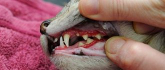
The use of aspirin or other NSAIDs is not uncommon during the history taking. Cases of ulceration and perforation of the gastrointestinal tract have been described in patients of veterinary clinics who received selective cyclooxygenase inhibitors in therapeutic doses. Decreased or lack of appetite in patients receiving NSAIDs should suggest possible complications from the gastrointestinal tract. The use of drugs should be discontinued and the patient examined. If the cause of gastrointestinal bleeding in cats and dogs is thrombocytopenia or coagulopathy, there may be a history of bleeding from other sites, including the nasal cavity or urinary tract. In patients with severe thrombocytopenia, petechiae can be detected on the mucous membranes when examined. Examination of subcutaneous nodules or tumors will identify underlying mastocytomas.
The abdomen (abdomen) should be carefully examined because bleeding from the gastrointestinal tract
may occur and develop asymptomatically, especially chronic bleeding. Palpation of the abdominal wall will allow you to localize the area of pain (weakness, a voluntary or involuntary attempt to defend yourself, or induce vomiting, identify tumors or foreign bodies, determine the enlargement of the abdomen (stretching of the abdominal wall) or signs of the presence of fluid in the abdominal cavity (waves during percussion). In patients with mastocytoma or other neoplasias, liver disease may show hepato or splenomegaly. At the initial examination or during resuscitation, a thorough rectal examination should be performed to identify melena or pure blood, tumors or foreign bodies. Although bleeding from any part of the gastrointestinal tract can be serious, bleeding from the upper tract tends to be more severe. In addition, the etiology, diagnostic techniques, and treatment of upper and lower GI bleeding are different. Thus, it is important to determine the area of bleeding. The presence of hematomesis or melena suggests upper GI bleeding. However, it should be remembered that blood remains in the gastrointestinal tract for some time and it is not necessarily this location that determines the color. Increased transit time and retention in the large intestine lead to the appearance of melena, which is associated with damage to the lower gastrointestinal tract. Hematochezia usually reflects bleeding from the colon, rectum, or anus. However, acute severe intestinal bleeding acts as a laxative, which significantly reduces the transit time. This may result in pure blood in the stool due to significant blood loss from the upper GI tract.
Diagnostics
Bleeding in cats and dogs has been proven
when the source of bleeding is identified. In patients with signs of shock, during correction of the condition and search for hidden causes, a minimum number of emergency blood tests are performed, namely hematocrit, solids, blood nitrogen (BUN), glucose, if possible, pH, lactate and electrolytes. If hemoabdomen or septic peritonitis is suspected, emergency ultrasound, laparocentesis and diagnostic peritoneal lavage are indicated, which can be performed during the initial correction of the patient's condition. Once resuscitation has been initiated or the patient's condition has been stabilized, other diagnostic modalities should be considered.
Treatment of peptic ulcer
Treatment for gastric ulcers in cats is symptomatic: water and electrolyte balance is restored, drugs with analgesic and antiemetic effects are prescribed, and a dietary regimen is prescribed.
If there are signs of dehydration, infusion therapy is performed. A dropper is installed, the basis of which is a 0.9% solution of sodium chloride with glucose or other components (as prescribed by the treating veterinarian).
If the development of a stomach ulcer is provoked by pathogenic microorganisms Helicobacter Pylori, antibacterial therapy is carried out, and drugs are prescribed to increase blood clotting. In severe cases, when the bleeding cannot be stopped, a gastrotomy is performed.
The next stage of treatment is the restoration of the cat’s body. In order to strengthen internal organs and systems, vitamins are prescribed:
- A (retinol),
- E (tocopherol),
- C (ascorbic acid),
- B6 (pyridoxine).
The key to successful treatment is the elimination of the factor that provoked the development of gastric ulcer. Upon completion of therapy, we recommend regularly visiting the veterinarian to eliminate the possibility of relapses.
Tests to Confirm Gastrointestinal Bleeding in Small Pets
If physical examination and history do not reveal obvious symptoms of gastrointestinal bleeding in dogs and cats, hematological and biochemical studies are performed, the results of which may suggest bleeding. Anemia of unknown etiology may lead to the assumption of bleeding from the gastrointestinal tract; it is described that microcytic hypochromic anemia (iron deficiency anemia) develops as a result of chronic blood loss in the gastrointestinal tract. Because iron deficiency anemia takes time to develop, a more typical consequence of recent bleeding is normocytic normochromic anemia. A high urea-creatinine ratio (greater than 20) has been described in gastric and intestinal bleeding, especially if the bleeding occurs from the upper gastrointestinal tract. This is due to bowel movements and intestinal absorption of proteins (may increase the level of digested and absorbed protein), including digested blood, into the circulatory system (blood). Massive intestinal bleeding, as described, has little effect on urea levels. However, once protein metabolism is activated as a result of illness (fever, infection, fasting or use of glucocorticosteroids), the urea-creatinine ratio may also increase, which must first be concluded to be the cause of gastrointestinal bleeding in dogs and cats.
In doubtful cases, a fecal occult blood test is performed. Most tests are based on the peroxidase activity of hemoglobin. Although the test is useful in detecting occult bleeding, it is possible to obtain a false positive result. This is influenced by the following factors: diets that include red meat or foods with high peroxidase activity, such as fruits, fish, vegetables. The result is also affected by bacteria with peroxidase activity present in the gastrointestinal tract. This must be kept in mind when receiving test results based on chemical characteristics.
It should be recommended to exclude meat from the animal’s diet for at least 72 hours. On the other hand, a negative occult blood test excludes significant GI bleeding. When significant gastric bleeding is suspected but not confirmed, a nasogastric tube is inserted and the gastric contents are aspirated. This will help confirm and determine the location of the bleeding. Although the procedure can cause discomfort and falsely described - negative results.
Classification of abdominal injuries in cats
Cats suffer from abdominal injuries such as:
- Penetrating wounds. Characterized by rupture of the peritoneal wall and adjacent internal organs. Bleeding begins and peritonitis quickly develops.
- Injury. It manifests itself as swelling, edema, hematoma, and soreness of muscle tissue. In this case, there is always a risk of organ rupture, which invariably leads to internal bleeding.
- Hemorrhage in the retroperitoneal space. Occurs as a result of closed injury to the kidney, liver, intestines, and stomach.
An open abdominal injury can be detected visually even by the owner of the animal. But bruises and hemorrhages can not always be recognized, especially if the cat is in a state of shock.
Tests to identify hidden causes
Once gastrointestinal bleeding in dogs and cats is suspected or confirmed, a search for an underlying cause should be undertaken: coagulation tests, complete blood count, routine chemistry profile, electrolytes, ACTH stimulation test, imaging (x-ray, ultrasound), endoscopy.
A coagulation test will help identify coagulopathies such as rodenticide poisoning or clotting factor deficiency. An increase in blood clotting time, which can be detected by testing, is not in itself the cause of gastrointestinal bleeding in cats and dogs, but can significantly aggravate the condition. The platelet count is important because a common cause of severe gastrointestinal bleeding in dogs is immune-mediated thrombocytopenia. An increase in hematocrit in patients with acute hemorrhagic and relatively normal protein concentrations is suggestive of hemorrhagic gastroenteritis.
Taking into account that the confirmed cause of ulceration of the gastrointestinal tract is liver and kidney diseases, special attention should be paid to the biochemical markers of these diseases (alkaline phosphatase, ALT, AST and bilirubin - for liver diseases; urea, creatinine, phosphorus - for kidney diseases). It is necessary to examine the level of electrolytes and perform an ACTH stimulation test, since the confirmed cause of GIB is hypoadrenocorticism. Research is carried out if no other causes of bleeding are found. A stool smear, culture, and test for parvovirus will confirm the suspicion of an infectious disease. Measuring gastrin levels is recommended if ulcers recur and do not respond to therapy.
Radiography will reveal foreign bodies, tumors, or free air in the peritoneal space. The cause of pneumoperitoneum can be assumed to be gastrointestinal perforation if the patient has not undergone abdominal surgery in the recent past. Although contrast radiography will reveal mucosal defects as the cause of gastrointestinal bleeding in cats and dogs, in most cases it can be replaced by ultrasound or endoscopic examination. Ultrasound can identify tumors and foreign objects accompanying gastrointestinal ulcers, if any. The use of ultrasound to recognize (define) ulcers is described. The study allows you to visualize the structure of the intestinal wall, its thickening, defects and craters. If serial testing is carried out (over time), it is possible to confirm changes that occur in response to therapy or the need for surgery in some cases. The use of ultrasound to identify ulcers in cats has also been described.
Endoscopy with greater accuracy (sensitivity) will allow you to determine the location of bleeding or ulcers in the upper gastrointestinal tract. Before the study, the patient's condition must be stabilized. The study often allows you to establish a diagnosis, assess the prognosis, can be therapeutically useful (removal of foreign bodies), and finally, during the study you can directly visualize the mucosa, obtain material for biopsy and culture, which may be necessary to determine damage or infection (for example, tumors, inflammatory bowel disease, protothecosis Disadvantages of endoscopy include the need for anesthesia, limitation to the proximal GI tract and colon, potential increased bleeding, and the possibility of iatrogenic ulceration.
If all of the above diagnostic methods do not allow us to establish the cause of ongoing gastrointestinal bleeding, then diagnostic laparotomy, scintiography with technetium-labeled erythrocytes and arteriography are performed. Using scintiography, you can determine the source of bleeding in dogs, and arteriography will allow you to establish vascular pathology of the gastrointestinal tract.
Treatment of dogs and cats with bleeding from the gastrointestinal tract
The primary goals of treatment are to stabilize the patient's hemodynamics, control ongoing bleeding, treat existing ulcers, prevent bacterial translocation, and address underlying causes. The initial priority is to immediately identify and treat any signs of shock.
Depending on the duration and extent of blood loss, a transfusion of red blood cells, whole blood, or oxyglobin may be required. For patients with acute severe bleeding, blood transfusion is indicated as part of the initial resuscitation measures. For patients without signs of shock, the indications for transfusion of blood products are less clearly defined. The decision to transfuse blood products for all patients is based on hematocrit values, which is still controversial. The hematocrit level at which patients require transfusions may vary depending on the degree of blood loss, hemodynamic status, initial and subsequent hematocrit measurements, the presence of comorbidities, and the severity of clinical signs. If the patient has signs of decreased oxygen delivery (tachycardia, hyperlactemia, tachypnea) or if serial measurements show a decrease in hematocrit after initial therapy, then a blood transfusion is indicated.
If gastrointestinal bleeding in dogs and cats develops as a result of a primary coagulopathy or is aggravated by a secondary coagulopathy (DIC, liver disease, shock, dilution due to aggressive fluid therapy), fresh frozen plasma transfusion should be considered. In patients with persistent gastrointestinal bleeding in animals due to thrombocytopenia, the use of vincristine, which increases the release of platelets from the bone marrow, may be beneficial. Although the functionality of these cells remains in question.
As a means to reduce bleeding from the digestive tract, you can use gastric lavage with cold saline solution. Although, according to the recommendations presented in the medical literature, this method should be avoided. Lavage has not been proven to be effective in slowing bleeding. But it is known that it can cause discomfort and lead to a rapid drop in body temperature, which can lead to prolongation of bleeding, which has been proven in experimental studies in dogs.
Animals with hematomesis and melena should receive antiulcer therapy until another cause of symptoms is proven. Drugs whose use may lead to ulceration should be discontinued. Given the association between the use of steroids in dogs and gastrointestinal bleeding in dogs and cats, these drugs should also be discontinued unless these drugs form the basis of therapy, for example, for hypoadrenocorticism, immune-mediated diseases.
It is reasonable to use gastroprotectors before confirming the diagnosis of gastrointestinal bleeding in dogs and cats, given that ulcers are the most common cause of EKC in dogs and cats, the use of gastroprotectors may have a wide margin of safety(?). In addition, intraluminal (intraluminal) neutralization of gastric acid may slow GI bleeding by maintaining mucosal homeostasis. Commonly used gastroprotectors include acid suppressors such as H-2 receptor antagonists (cimetidine, famotidine, ranitidine) or proton pump inhibitors (omeprazole, pantoprazole), mucosal binders such as sucralfate and synthetic prostaglandins (misoprostol). There is no veterinary research data confirming that the use of gastroprotectors or their combinations are more effective in the treatment of gastrointestinal ulcers. However, studies demonstrate that famotidine (0.5 µg/kg every 12 hours), omeprazole (1 mg/kg every 24 hours PO) and pantoprazole (1 mg/kg IV every 24 hours) significantly suppress gastric acid secretion in dogs, but ranitidine (2 mg/kg IV every 24 hours) is not able to significantly suppress gastric acid secretion when the dose is increased.
Although H-2 receptor antagonists, proton pump blockers, and sucralfate are used together, in most cases of suspected gastrointestinal ulcer, either H-2 receptor antagonists or proton pump blockers and sucralfate are used. In case of poisoning (intoxication) with NSAIDs, misoprostol can provide an additional positive effect. When choosing a drug, it is necessary to base it on the method of its use, since the absorption of drugs from the gastrointestinal tract (when administered orally) in critically ill patients is questionable. Most dogs with gastrointestinal tract vomiting present, which limits further oral administration of drugs and their disposal. In patients with persistent vomiting, the use of antiemetics is indicated. Metoclopramide (1–2 mg/kg every 24 hours) is often used for initial therapy. If vomiting is refractory to metoclopramide, ondansetron may be helpful. Because gastrointestinal bleeding in cats and dogs is often accompanied by discomfort and pain, analgesics such as opioids should be used.
If bleeding is significant, use of broad-spectrum antibiotics (eg, penicillin and an aminoglycoside or fluoroquinolone, or a combination of cephalosporins, metronidazole and an aminoglycoside or fluoroquinolone) is indicated. This is due to the fact that with gastrointestinal tract infection the gastrointestinal tract barrier is disrupted and there is a possibility of bacterial translocation. Ideally, cultures and susceptibility testing (eg, blood and urine) should be obtained before starting antibiotic therapy.
For the most part, gastrointestinal bleeding in dogs and cats is amenable to therapeutic correction. If the ulcer or gastrointestinal tract is refractory to therapeutic treatment, endoscopic hemostasis may be useful. Hemostasis of ulcers(?) has been described by injection of epinephrine or 98% alcohol through an endoscopic sclerotomy needle into the base of the ulcer. The use of minimally invasive thermal, laser, or electrical cautery or the use of an endoclip has been described to control intestinal and gastric bleeding secondary to vascular disease and ulcers in humans. These methods can also be used in veterinary medicine. Surgery can be avoided in many cases, but is indicated for existing surgical pathologies (foreign bodies, tumors, septic abdomen), in patients at risk of bleeding or perforation (based on endoscopy or serial ultrasound), or if the patient is not responding to therapeutic treatment.
Since many pathological conditions lead to the development of gastrointestinal bleeding, therapy aimed at correcting the underlying disease can be different (for example, surgical removal of a foreign body or tumor, steroids for hypoadrenocorticism, immunosuppressive therapy for immune-mediated thrombocytopenia, withdrawal of NSAIDs). Given occult disease, it is important to evaluate for associated or unrelated coagulopathy (eg, liver disease leading to ulceration and deficiency of thrombus-forming factors) and to look for concomitant disease that may aggravate GI bleeding in dogs and cats (eg, uremia in patients using NSAIDs). See Absorbing agents
Features of internal bleeding
Internal bleeding is the release of blood from damaged vessels into the body: into the abdominal or pleural cavity, into the space between muscles, into subcutaneous tissue, as well as inside hollow organs, for example, into the bladder, trachea or stomach.
The degree of danger to the pet's life depends on the rate of bleeding. With chronic, relatively small blood losses, oxygen starvation of the body tissues begins, anemia develops, metabolic processes are disrupted, which entails cell death. The animal’s activity decreases, the quality of its fur deteriorates, and the pet loses weight to the point of anorexia. In rare cases, lethargy develops.
Bleeding may stop due to the formation of clots that block the breakthrough, and then resume again due to their displacement. In this case, periods of weakness in the animal will alternate with normal health.
A blood clot can block a damaged vessel, but this improvement will only be temporary
With severe internal bleeding, the cat can die both due to a drop in pressure and oxygen starvation, and due to blood squeezing vital organs, such as the heart or brain. Without qualified assistance, death in this case occurs within a few hours or minutes, depending on the type of injury.
Good to know
- Esophagoscopy in dogs and cats
- Protein wasting intestinal disease in dogs. Enteropathy in dogs
- Treatment of hyperlipidemia in dogs with Bezafibrate
- Vomiting, regurgitation in dogs and cats
- Small intestinal transient disorders
- Diseases of the anus and rectum in dogs and cats
- Cholangitis in cats. Identifying, diagnosing and treating cats with neutrophilic, bacterial, lymphocytic or chronic cholangitis
- Diarrhea in dogs and cats
- Hematemesis in dogs and cats
- What are the main causes of protein-losing enteropathy in dogs?
- Main causes of chronic colonic diarrhea in dogs and cats
- How to distinguish chronic small intestinal diarrhea from large intestinal diarrhea?
- Causes of malabsorption in dogs
- Vomiting in cats and dogs
- Causes of acute diarrhea in dogs and cats
- Canine distemper in dogs (forms, diagnosis, symptoms, treatment, therapy)
- Canine exocrine pancreatic insufficiency
- Enteral nutrition for dogs and cats
- Protein-losing enteropathy. Protein enteropathy in dogs
- Lymphocytic cholangitis in cats
- Acute pancreatitis in dogs and cats
- Gastric volvulus in dogs
- Fat metabolism and hyperlipidemia in dogs