Formation of primary dental occlusion
Almost all mammals are born toothless. Cats are no exception: the oral apparatus of a newborn kitten is maximally adapted for effective sucking. After all, the only food available to him is mother’s milk.
At two weeks of age, kittens develop milk teeth - straight, small, sharp, milky white. In adult cats they are powerful and curved. They are called milk teeth because they appear in kittens during breastfeeding.
Interesting! The front milk teeth of kittens are straight, and the fangs are curved.
Teething order
The eruption period lasts up to two months. By this age, the cat already has 26 baby teeth. They do not appear all at once, but in a certain order:
- 2–4 weeks. Incisors appear. First, 2 in the center of the jaw, then 4 more are added to them on both sides. There are 12 incisors in total - 6 each on top and bottom.
- 3–4 weeks. Fangs appear: 4 upper and 4 lower. They are located on the sides of the incisors.
- 4–8 weeks. The appearance of pre-masticating teeth - premolars: 4 on the lower jaw and 6 on the upper jaw.
Important! Milk teeth will serve kittens up to the age of 3-4 months with proper care and timely introduction of complementary foods.
Formation of teeth in a kitten
The structure of the teeth of cats is typical for their species, and zoologists consider the structure of the dental system as its morphological feature. The structure of the dental system reflects the nature of the diet, and the cat is an undisputed predator - all its dental crowns are conical in shape and are designed to tear hunted game into pieces. The teeth located on the jaw form the upper and lower arcades, respectively. To form a breed characteristic—the appearance of a cat’s face—the correct formation of dental arcades is necessary. Teeth are necessary for capturing, holding and tearing prey; the cat uses them to intimidate the enemy, in fights, for grooming, and for carrying small objects, including kittens, in its mouth.
The formation of tooth buds in cats occurs during embryonic development in the form of a dental plate - this is a fold of epithelium containing tooth buds. The inner layer of cells of the dental plate - anameloblasts, form tooth enamel; The cells of the outer layer are odontoblasts, they form dentin. The connective tissue surrounding the tooth germ forms cement.
The development of teeth is associated with their eruption, when the teeth come out through the mucous membrane of the gums.
Teeth are made up of tissues:
- dentin is the modified bone tissue that makes up most of the tooth. Dentin forms the tooth cavity, where the pulp containing blood vessels and nerve fibers is located;
- enamel is a very durable white tissue that covers the surface of the tooth from the outside, where it comes into contact with the external environment;
- cement - covers the parts of the tooth located in its alveolus - the cell of the jaw where the tooth is located. Cementum is represented by bone tissue of a layered structure.
The following parts can be distinguished in the appearance of a tooth:
- tooth crown - part of the tooth that protrudes freely into the oral cavity and has a chewing surface;
- tooth root - located in the alveolus; the cementum of the tooth root and the periosteum of the alveoli are connected by the periodontium;
- neck of the tooth - connects the crown of the tooth with its root, here the coating of the tooth changes, the enamel of the crown passes into the cement of the tooth root. The neck of the tooth is normally located in the gum.
Based on function and form, they are distinguished:
- incisors - small, flat-shaped teeth located on the jaws in front and having one root. The incisor crown has three projections that wear away over time; The cat uses the incisors for grasping and holding, as well as grooming; the roots of the incisors are shallowly embedded in the jaw, so these teeth are often lost with age;
- fangs - the cat’s fangs are dagger-shaped, they are sharp and long, their roots are immersed very deeply in the bone tissue of the jaws; used to kill prey; the cat uses its fangs to separate the cervical vertebrae of the victim - this is a characteristic hunting feature of small cats; fangs are used for tearing food; each fang has only one root, the fangs are located next to the incisors;
- premolars - small molars; Kittens also have them; premolars are needed for grinding food and have a chopping effect; the number of premolars is different on the upper and lower jaws: there are 6 on top, 4 on the bottom. They have 2 or 3 roots, so before removing a premolar, the veterinarian prescribes an x-ray to clarify the number of roots and not leave one of them in the jaw; located next to the fangs;
- molars - large molars - do not have their milk equivalent, they grow immediately in the form of permanent teeth; molars are also designed for cutting and crushing food; located at the edges of the dental arcades; The upper molars have one root each, the lower molars have 2.
During the teething period, cats try everything, it is especially dangerous when they chew wires
When and in what quantity do baby teeth appear?
At birth, a kitten has no teeth at all, but starting from the 2nd week of life, milk teeth begin to appear. A cat's primary set of teeth is incomplete because molars, the large molars, are missing. They are called milk teeth because they appear when the kitten feeds on milk, and also because of the specific color of the tooth enamel - milky white, translucent. Baby teeth are smaller, sharper and more fragile compared to permanent teeth. In total, the milk set contains 26 teeth: 12 incisors, 4 canines, 10 premolars.
Timing for the eruption of baby teeth:
- incisors: the first primary incisor erupts at 2–3 weeks; the second - at 2.5–4 weeks; third - at 3–4 weeks;
- fangs: milk fangs erupt at 3–4 weeks of a kitten’s life;
- premolars: eruption in 4 to 8 weeks.
It is generally accepted that the age of a kitten can be accurately determined by its teeth, but this is not entirely true, since individual and breed characteristics of the timing of teething can create a large error in the accuracy of this method.
The practical benefit of knowing the average timing of the eruption of baby teeth is expressed in the ability to control their development, since the development of teeth is also an indicator of the development of the kitten.
The eruption of baby teeth usually goes unnoticed by the kitten’s owner, because the cat is still too small.
By 2 months the kitten has a full set of baby teeth.
At what age does teeth change?
The replacement of baby teeth with permanent ones occurs when the kitten is 3–6 months old. The permanent set includes 30 teeth: 12 incisors, 4 canines, 10 premolars, 4 molars.
Changing baby teeth to permanent ones
The replacement of baby teeth with permanent teeth in cats begins at 4 months. The formation of the masticatory apparatus is completed at 7–8 months. The time it takes for a permanent bite to develop may vary among different cat breeds. A slight advance or delay in deadlines is allowed.
Shift order
The location and order of changing baby teeth to permanent ones is clearly depicted in the figure below. The first to fall out are the deciduous incisors. Fangs change after complete replacement of incisors. Next comes the replacement of the premolars. After a complete change, 4 molars erupt - the main chewing ones. In the picture they are colored blue.
Types of permanent teeth
An adult cat should have a total of 30 teeth. For a cat, it is a weapon, a means of protection, defense and attack. Mother cats use their front incisors to gently grab and carry the kitten.
Interesting! In cats, the lower jaw can only move in a vertical plane, so it is incorrect to say that cats chew their food. They crush it, crush it into small pieces.
The table describes the location of cat teeth, their number and functional load.
| Upper jaw | Incisors – 6 | They serve to hold prey and tear off small pieces of food. | Fangs – 2 | Used for self-defense, capturing and chopping food | Premolars – 6 Molars – 2 | They grind the bones and crush large pieces. |
| Lower jaw | Incisors – 6 | Fangs – 2 | Premolars – 4 Molars – 2 |
Molars are located deep in the jaw. Their presence indicates that the cat has become an adult. During this period, he begins to mark his territory.
Symptoms of teething or changing teeth
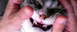
By the behavior of kittens and the reaction of a nursing cat, you can understand that they are cutting their first teeth. The appearance of primary incisors is accompanied by swelling and soreness of the gums. The kitten is trying to get rid of unpleasant sensations, so during feeding it clenches its jaws with force and bites its mother's nipple.
Sharp incisors can injure the nurse. But this period does not last long and often goes unnoticed by the owners.
By the end of the 4th month, the rudiment of a permanent tooth is formed in the gums of the upper and lower jaws under each milk tooth. As it grows, baby teeth shift, are pushed out of the gums and fall out one by one. This occurs at approximately 5 months.
Replacing baby teeth with permanent ones causes noticeable changes in the kitten's behavior. During this time, your pet's condition should be monitored to help if complications arise.
Symptoms of teeth changing in kittens:
- When young cats lose their baby teeth, they become biters in an attempt to get rid of itchy gums. You should not allow the kitten to bite the owner's hands. It will be very difficult to re-educate him later.
- Excessive salivation appears - ptyalism. This is how nature protects delicate gums from possible inflammation.
- An unpleasant odor appears from the mouth. The reason is moderate inflammation.
- Possible increased body temperature, lethargy or agitation, aggression. This is a reason to contact a veterinarian.
- Kittens lose their appetite or refuse solid food because their swollen, inflamed gums are painful to chew. Dry food should be removed from the diet, leaving only soft food.
Important! When kittens change teeth, it is necessary to exclude the animal from access to anything that could harm it. For example, when chewing wooden surfaces, a cat can hurt its gums.
To make the teething process go smoothly, you can purchase special “chewing” toys.
Lost primary incisors can be discovered by the owner during play. But most often kittens swallow them while eating. This is normal - everything will work out naturally.
Possible complications when changing teeth
Changing teeth in a kitten is a natural process. It can go smoothly, or it can get complicated.
Mild inflammation or redness of the gums is normal. However, it is worth observing all changes and, at the slightest suspicion of deviations, contact a veterinarian.
Symptoms that indicate possible problems:
- suppuration forms on the wound from a lost tooth;
- the kitten is in a bad mood, lethargic, restless;
- refusal to eat for more than a day;
- gums are very inflamed;
- formation of wounds near grown molars;
- The permanent teeth have grown, but the baby teeth have not yet fallen out.
The appearance of any of these signs should be a reason to contact a veterinarian.
Other problems
It happens that the entire row of permanent teeth has formed, but the baby teeth have not fallen out. This situation causes jaw injuries, crooked bites, and the development of periodontal disease. A qualified doctor will help you deal with this problem.
A very common problem when changing teeth is improper growth of fangs. There are several variants of this pathology, so the problem is corrected individually in each case. But this cannot be ignored, since improper tooth growth worsens the animal’s quality of life and creates digestive problems.
Each case of abnormal formation of fangs in a cat is considered individually
Possible complications
The eruption of baby teeth and their replacement with permanent teeth are natural processes of growth for any animal. They must take place on time. It is necessary to contact a veterinarian in time if complications arise:
- The kitten has been sleeping poorly for the last 24 hours, not eating, restless, and meowing.
- The unpleasant odor does not go away for a long time, it becomes pungent - periodontal disease may develop.
- Excessive secretion of saliva and its thickening - the likelihood of gingivitis or stomatitis.
In order not to miss alarming symptoms when cats change teeth, you need to regularly examine the oral cavity.
Gum inflammation
A healthy cat's gums should be pale pink and free of swelling. During eruption, swelling, redness, slight swelling and short-term bleeding are allowed. These phenomena quickly pass: the swelling quickly subsides, the hole in the place of the lost tooth heals without a trace.
You should not postpone a visit to the veterinarian if:
- inflammation has occurred around the baby tooth;
- Reddened gums bleed for a long time;
- pus appears on the gums or in the sockets;
- the baby incisor is “slanted” and injures the gums, this usually happens with fangs.
All of these signs are symptoms of periodontal disease, stomatitis, and gingivitis. Purulent inflammations are especially dangerous.
Interesting! Scottish Fold cats are the most prone to gum disease. The owners of Scottish dogs should be especially attentive to them during this difficult period.
Residual baby teeth
Usually the new tooth grows in its own socket. Therefore, it often happens that a permanent one grows next to a milky one that has not yet fallen out. This is the norm, but if a baby tooth does not fall out in 8 months, this is a reason to consult a doctor.
An anomaly, when permanent teeth have already erupted, but milk teeth have not yet fallen out, is rare. In this case, a double dentition is formed and, as a result, an incorrect bite. In this case, the oral mucosa, the inside of the cheeks, and the kitten’s tongue are injured.
If more than one or two primary incisors remain in the pet's jaw, surgical intervention and strict monitoring of the subsequent eruption process are inevitable.
Expert opinion
Chepa Natalya Semenovna
Veterinarian
Ask an expert
In some cases, kittens may develop juvenile gingivostomatitis after teething. This is an inflammation of the gums that develops after the appearance of permanent teeth. Young cats under 2 years of age are affected. The exact causes of the inflammatory process have not been fully established. The disease probably occurs against the background of an allergic reaction of the body to plaque bacteria. Some breeds have a predisposition: Maine Coons, Sphynxes, Bengals, Orientals. Characteristic signs of juvenile gingivostomatitis are bad breath, redness and swelling of the gums. To treat the disease, sanitation of the oral cavity with antiseptics, for example, Miraxidin solution, is prescribed. In difficult cases, ultrasonic teeth cleaning may be required. To prevent the disease, it is recommended to brush your pet’s teeth with special pastes for animals. Moreover, it is better to accustom your cat to the procedure from childhood.
Correction of canine position disorders in dogs and cats
F. Genne
Violation of the position of the canines is a change in the bite, which affects their functional state.
Incorrect canine alignment can be caused by a misalignment of the primary teeth or an abnormality in the size of the mandible.
The categories of these deviations from the norm are determined by the orientation of the canines in space.
The first part of the article presents a classification of violations of the position of the canines, while the second discusses the issue of ways to eliminate defects.
Classification
Abnormalities in the position of the teeth In the transverse projection An anomaly in the position of the fangs is much more often noted in the transverse projection. It is mainly found at the level of the lower canines, the position of which has a pronounced direction towards the tongue.
This anomaly, called canine convergence, refers to linguoversion (severe inclination of the canines towards the tongue) or linguoposition (severe displacement of dental implants towards the tongue). The etiology of these dental disorders is not well understood. If in small breeds of animals the persistence of primary canines is the main factor of these disorders, then in others this cause of defect is encountered much less frequently. Also, the lower canines may have a pronounced inclination in the lateral direction (vestibular version), which is also called divergence.
As for the upper canines, violation of their position is much less common. We can observe their deviation towards the palate (due to the persistence of baby teeth).
In lateral projection In dogs and cats, rostral deviation (rostroversion) of the upper canines can also be observed. This is most often observed in small breeds of dogs (Yorkshire terrier, poodle, dwarf dachshund, etc.) due to the persistence of primary canines. In addition, depending on the breed, the persistence of primary canines causes a more or less pronounced disturbance in the position of the permanent canines. This feature is typical for Italian and Scottish Italian greyhounds (photo 1). In any case, particularly in these breeds, it is very difficult to establish whether the persistence of the primary canine is the cause of this type of disorder or whether it provokes rostroversion of the primary canine.
| Photo 1. Rostroversion of the upper canine in an Italian greyhound. A persistent milk tooth was recently removed (the wound caudal to the tooth), but a fragment remained. |
In cats of brachycephalic breeds (Persians, exotics), these disorders are of a specific nature. If there is rostrodeviation of the upper canines in association with the persistence of primary teeth, then it should be borne in mind that one of the reasons for the intensity of this pathology is the maxillary-facial modification that we observe in brachycephalic breeds. As for the lower canines, the violation of their position is caused by the rostral deviation of the upper canine or a skeletal anomaly, characteristic of brachycephals.
Vertical projection A fairly common anomaly in the position of the canines in the vertical projection is egression (progression of one or more teeth that do not have antagonists), and it is observed mainly in small breeds of dogs. Egression is also noted with malocclusion.
Violation of the position of the teeth associated with changes in the structure of the skeleton In the transverse projection As for the violation of the position of the main teeth at the level of the lower canines (“convergent canines”), it is recommended to introduce the term mandibular endognathium (narrowing of the lower jaw). This skeletal anomaly in transverse view corresponds to a pronounced "internal" displacement of the position of the canines in relation to the maxilla.
In the lateral projection In the case of upper or lower prognathism (or brachygnathism), a violation of the position of the upper canines in relation to the lower canines is noted, due to the shortening of one of the jaws.
These two anomalies are regrouped because they are predominantly volumetric anomalies (transverse and sagittal projection), in which the mandible is narrower or shorter (micromandibularia). In this case, the lower canines have a more internal and caudal position.
Guide to action
Reason for consultation and medical history There are two reasons for consulting a doctor:
- mainly rostral deviation or divergence of the lower canines (the aesthetic factor plays a role here);
- indications for treatment in case of anomaly of the lower canines in the transverse projection (convergent canines) or in the case of rostroversion of the upper canines or micromandibulia.
The main causative factors that must be taken into account are: breed, how long ago the anomaly occurred, the state of occlusion due to the replacement or spontaneous loss of primary teeth, the presence of primary teeth, as well as the presence of anomalies during the growth period.
Determining the severity of the violation The main significance is the violation of the position of the lower canines. Due to their size, changes in the position of these teeth, mainly in the transverse projection, negatively affect the condition of the gums or palate. Sometimes the disease develops before the change of baby teeth, but becomes more pronounced from the moment the permanent teeth appear (from the age of five months). Treatment should be carried out before the age of one year (photo 5, 6).
| Photo 5. Damage to the palate due to linguoversion of the lower canine in a puppy aged six months. It should be noted that the canines have not yet fully erupted. | Photo 6. Occlusion of the maxillary teeth in this dog indicates damage to the palate, which is localized in the space between the teeth. Effective orthodontic treatment was carried out (Fig. 1). The lips fit tightly to the upper canine, which ensures fixation of the removable appliance. |
| Figure 1. Treatment options for palatal injuries caused by mandibular canine malalignment. 1) – damage to the palate is noted in the sagittal plane in relation to the space between the angle of the canine and the first third of its width. Orthodontic treatment is required. 2) – damage to the palate, located in the sagittal projection at the level of the second third of the width of the canine. It is necessary to amputate the crown. 3) – damage to the palate, localized in the sagittal projection at the level of the third third of the width of the canine. It is necessary to carry out amputation of the tooth crown or orthodontic treatment aimed at repositioning the tooth caudal to the upper canine, the tooth should be located between this canine and the first premolar |
Rostral deviation of the upper canines provokes a more or less complete closure of the space between the teeth (forming the shape of an angle) into which the lower canines lie. Consequently, the lower canine, subject as a result of the above changes to a certain degree of lateral (vestibuloversion) and/or rostral (rostroversion) deviation, provokes injury to the upper lip or covers it.
Making a diagnosis At the beginning of the consultation, it is necessary to carry out a differential diagnosis, if possible, between skeletal anomalies and a violation of the position of the tooth.
An external clinical examination of the patient allows one to assess the degree of symmetry of the skull and facial surface of the head, as well as the position and shape of the main elements of the skeleton.
An oral cavity examination allows you to evaluate:
• occlusion of incisors, canines and premolars; • position of premolars (overfilling, rotation or displacement); • the shape of the mandible (straightness and parallelism in terms of occlusion or curvature with an open bite or gaping at the premolar level).
Before starting treatment, it is necessary to very carefully and completely examine the oral cavity under general anesthesia, which also makes it possible to conduct radiographic examinations inside the oral cavity. Radiography makes it possible to identify remaining fragments of the roots of primary teeth (photo 2), as well as to assess the stage of development of the roots of the corresponding teeth.
| Photo 2. Rostroversion of the upper canine in an Italian greyhound at the age of six months. The tooth is immature: the pulp canal is very wide, the root wall is thin, and the apex is unformed. It should be noted the persistence of a fragment of the root of a primary tooth (arrow). This persistence distorts the position of the main tooth. |
Treatment
Lower canines Lingual deviation of the lower canines Lingual deviation of the lower canines is most often accompanied by disturbances in the position of the teeth (linguoversion or linguoposition), the orthodontic treatment of which is quite well developed.
If baby teeth are still present, it is recommended to remove them immediately with care so as not to damage the root. In case of doubt, X-rays are used.
The decision for orthodontic treatment can be made when the lower canines have grown to half their height, which corresponds to the animal's age of approximately six months. The therapeutic option (choice), depending on the situation, is to install an active appliance between the lower canines or a passive one, which is also called functional, used on the upper jaw (at an angle).
If treatment is carried out before the full growth of the canines (at the age of 7 months), it is recommended to use:
• active flexible, usually hinged apparatus, so as not to change the direction of divergent growth of the lower canines; • functional appliance (angled), removable, designed for the upper jaw, which does not put pressure on the lower canines.
In addition, the transverse growth of the maxilla should not be inhibited during this period, which requires the use of an inclined hinged or “telescopic” appliance that meets these requirements (photo 7, 8). In adults it can be used with a fixed inclination.
| Photo 7. Installation of a functional removable appliance (inclined). The device is held passively using the lips. | Photo 8. The result of orthodontic correction after removal of the apparatus. |
Lingual and caudal deviation of the lower canines • Orthodontic treatment If the anomaly seen in the sagittal projection (caudal deviation) is not so pronounced, then a functional appliance (at an angle) can be used to correct the tooth(s) while simultaneously performing rostroversion (moving forward) or vestibuloversion (lateral displacement). During the examination, we can determine the boundary of the first third of the length of the upper canine (photo 6). The lower canines, which extend beyond its limits when the jaws are clenched (Figure 1, Photo 8), should be treated primarily in a different way: with the help of an active, more complex orthodontic apparatus (Photos 9, 10) or by amputation of the crown.
| Photo 9. Damage to the palate in the sagittal plane, at the level of the second third of the width of the canine. The most adapted treatment is crown amputation | Photo 10. Linguoversion and distoposition (more caudal position) of the lower canine. |
• Crown amputation Endodontic therapy is a priority when orthodontic treatment is contraindicated or undesirable. It allows you to avoid injury to the palate caused by the fangs if they are incorrectly positioned (photo 4, 8). This method of treatment is resorted to when the animals are over seven months old, because this corresponds to the period of formation of secondary, that is, more durable dentin at the crown level. This treatment is based on amputation of the upper part of the crown (approximately half). The tooth is then subjected to partial devitalization by eliminating the pulp (pulpotomy or partial pulpectomy). As a result of the manipulation, the formation of secondary dentin in the tooth crown is suspended. And vice versa, in its radicular (root) part the pulp is preserved: for this purpose we place a biological coating (“pulp combing”), which allows us to maintain its vital activity (normal root maturation) and in particular the closure of the apex (apexogenesis) and thickening of the root part of the wall tooth (secondary dentinogenesis). X-ray control for an objective assessment of its physiological formation is carried out after four or six months (photo 11, 12).
| Photo 11. Correction carried out by installing the maxillary and mandibular active apparatus. | Photo 12. Amputation of the crown with pulpotomy and “combing” of the pulp in a puppy at the age of seven months. The root is not yet ripe. Pulp brushing material and obturation are recognized by detecting a radiopaque mass. |
Rostral and vestibular deviation of the lower canines In case of deviation of one or two lower canines outward (lateral) and forward (rostral), the upper lip is captured, which is the reason for consultation. If the deviation is mild, then the canine(s) may rest against the upper lip.
This positional imbalance, either unilateral or bilateral, is common in brachycephalic cat breeds in association with skeletal disharmony. In this case, orthodontic treatment is not recommended, since the disorder occurs not at the level of the teeth, but at the skeleton. In cases where a violation of the position of the teeth is not associated with a skeletal abnormality, it is often unilateral (one-sided) in nature. The lower canine may be in a state of rostroversion due to that of the upper canine or an incorrect inclination of the tooth.
Orthodontic correction of canine alignment disorders involves the use of an active device that has an elastic module. Anchoring should be performed at least at the level of the lower premolars and molars to ensure secure anchorage.
Upper canines Rostral deviation In cases of severe rostroversion of the upper canines, the therapeutic choice is focused on tooth extraction or orthodontic treatment.
Treatment of this tooth pathology cannot be neglected, since the lack of its functional activity predisposes to the development of periodontal disease.
Due to the fact that this anomaly is mainly found in small breeds of dogs, this already indicates a high predisposition to the development of periodontal disease in them. Orthodontic treatment involves moving the upper canine back. This manipulation, due to the large volume of the root and, therefore, reliable fixation of the canine, requires a delicate approach.
Another difficulty is that the lower canine is in the path of the upper canine when it is displaced back and, if the anomaly is pronounced, then the visible part of the crown of the upper canine is very limited in size (photo 1). Finally, to ensure stability of the anchoring, multiple teeth must be involved. In this case, the active apparatus may consist of several buckles and/or elastic modules (photo 13, 14). The force of the elastic modules is adjusted using an orthodontic dynamometer.
| Photo 13. Radiographic follow-up performed 6 months later in a dog that had previously undergone crown amputation. Root maturation (closing of the apex and thickening of the root wall) indicates a favorable outcome of treatment, which preserved the viability of the pulp. | Photo 14. Maxillary active appliance, supplemented with an exiting buckle, designed for caudal displacement of the upper canine in a Chihuahua. |
An approximate standard when working with a cat is the force it produces, which is approximately 150 g (photo 15). The found normal occlusion of the upper canine relative to the caudal aspect of the lower canine may reduce the phase of natural retention provided by the interdigitation (cohesion) of these teeth.
| Photo 15. Active device, including elastic modules, used to distalize (caudally shift or lower) the upper canine in a cat. Simultaneous anchoring of the upper and lower jaws. |
Root maturation is regularly monitored using radiographic examination, which makes it possible to ensure the quality of orthodontic treatment, which does not have a negative effect on the pulp and root of the tooth (photo 3, 4).
| Photo 3. Orthodontic treatment of an Italian greyhound suffering from rostroversion. It should be noted that the apex of the tooth has not yet matured. | Photo 4. Control carried out after treatment. The appliance is removed after the tooth has been restored to normal occlusion, and attention should be paid to root maturation with closure of the canine apex, indicating that pulp vitality is preserved. |
Hygiene of the vestibule of the oral cavity and teeth is very important, and during the treatment period it must be carried out regularly. For this purpose, chlorhexidine gel is used, which is used daily by applying it to the teeth and gums.
Deviation of the palate, causing a violation of the position of the upper canines Deviation of the palate, causing a violation of the position of the upper canines, is an extremely rare anomaly in brachycephalic breeds of cats. It occurs due to the persistence of primary fangs. These teeth often cause aggression.
Malocclusion can be corrected very early orthodontically from the moment the canine reaches one third of its height. In this case, an active mini-device in the form of an elastic buckle is used, which makes it possible to tilt the upper canine in the lateral direction (photo 16). Treatment does not always allow achieving complete egression of the tooth.
| Photo 16. Tensioning the elastic chain using a dynamometer. The tension is adjusted to a weight value of approximately 150 g. | Photo 17. Maxillary active appliance, complemented by a buckle, designed to bring the upper canine laterally (to the position of vestibuloversion), arriving at the moment in a state of palatoversion. |
Conclusion
Violations of the position of the canines are common and are the cause of a violation of their occlusion, which is responsible for the disruption of their functional activity, which should not be neglected.
The clinician must offer adequate treatment. Scientific knowledge and its practical application in the treatment of animals are constantly evolving in veterinary medicine. Lack of treatment or tooth extraction is currently not the only solution to this problem. The veterinarian, depending on the wishes of the animal owner and his capabilities, either independently chooses the method of treatment or consults with his colleague.
SVM 4/2005
Rate material
Like Like Congratulations Sympathy Outrageous Funny Thoughtful No words
Tags
Odontostomatology Orthodontic system Bite
Oral care
To maintain healthy teeth for your pets, you need to follow three basic rules:
- From an early age, it is necessary to provide the kitten with a healthy, balanced diet, sufficient amounts of minerals and vitamins. The veterinarian will help you choose food in accordance with the age and physiological characteristics of the cat. Cleaning of the incisors occurs naturally: when the animal chews solid food. That’s why the presence of dry food and large pieces in a cat’s diet is so important. Mechanical action on the surface of the teeth cleanses them of plaque and protects them from the formation of tartar.
- Compliance with the rules of basic oral hygiene. The list of kitten care procedures should include mandatory cleaning. Baby teeth can be cleaned with a soft cloth. The kitten will enjoy this procedure if a special paste for cats is applied to the fabric - it has a pleasant smell and taste. Changing incisors can be cleaned with a brush: the kitten will happily chew on the soft bristles, which will help cope with the itching. As your permanent teeth grow in, you can move on to brushing with a stiffer bristled brush. The procedure is carried out with light movements from roots to ends. Regularity – 2–3 times a week.
- Constant availability of sufficient clean water.
If tartar or other pathologies appear, you should contact your veterinarian. The treatment takes place under anesthesia, so there is no need to worry about the moral and emotional state of your pet.
Self-examinations conducted by the owner and regular visits to the veterinarian will help raise a strong, beautiful pet and maintain the health of its teeth for a long time. Regularity is important - this will allow you to identify problems in time and help the animal.
Determining the age of an animal by the condition of its teeth
Based on how the teeth look and their number, a specialist can fairly accurately determine the age of a cat. This may be necessary if the animal is picked up on the street. The condition depends on the influence of external and internal factors, the quality of food consumed. Therefore, age determination from them is made only approximately - with an accuracy of up to six months.
Here are the main signs by which one can judge age:
- the presence of 30 pieces of pure white color indicates an age of about a year;
- by 1.5 years of age, the teeth become slightly yellow;
- by the age of 2 years, the animal’s lower incisors are slightly worn, yellowness is clearly visible on the upper ones;
- by the age of 3, the process of abrasion of the upper incisors begins;
- by the age of five, pets’ incisors begin to change: they slightly change shape due to abrasion, becoming more yellow;
- incisors may fall out or become significantly weakened by age 7-8;
- By the age of 10, a cat may lose its fangs.
The incisors may fall out completely by the age of 12-14, followed by the fangs by the age of 15. This is not necessary and greatly depends on care, quality of nutrition, and the presence or absence of diseases of the oral cavity and dental tissue.
Cats' teeth should be properly cared for: feed the animal adequately, and regularly clean it using special pastes and brushes. The earlier in age you accustom your pet to grooming procedures, the more calmly he will tolerate them.
1111