Since childhood, we have heard “if you touched the cat, wash your hands.” If you have a pet, veterinarians constantly scare you: “Have you treated it for helminths?” That's it - anthelmintic treatment is required 10 days before vaccination.
Indeed, the prevalence of helminthiasis among cats is enormous, and the infestation does not depend on the living conditions of the animal - apartment or village. One only has to evaluate the incidence of helminths in people to understand the scale of the problem. Back in the 60s of the 20th century, Le Riche argued: for every inhabitant of Africa there are on average more than two types of helminths, in Asia and Latin America - more than one species, in Europe every third inhabitant is affected. According to official data, at least 15 million people in the Russian Federation are infected with helminths every year.
We humans have contact with helminth eggs absolutely everywhere - public transport, staircase railings, work in the country, in the garden, public places, etc. Cats that have free access to walks become infected just like humans, plus the risk of infection increases due to hunting mice, rats, and birds. “Apartment” cats become infected from us, since we carry a large number of eggs on shoes, clothes, bags, etc. For this reason, to prevent the development of helminthic infestation in cats, it is recommended to treat the animal for helminths quarterly (once every 3 months).
What nematode worms look like: photo and description
Many roundworms live freely exclusively in the external environment: soil, water bodies, animal bedding. Some types of roundworms parasitize plants (root-knot nematode, etc.).
According to the description, nematodes are worms with an elongated body of a spindle-shaped or cylindrical shape. Its cross section is round. Nematodes vary in size. These helminths are dioecious.
On the outside, the body of nematodes is covered with a dense cuticle, on which spines, ridges, papillae and other formations are visible. Under the cuticle there is a thin epithelial layer and well-developed muscles that form the skin-muscular sac. This bag contains the organs of the nervous, reproductive, digestive and other systems.
The role of fixation organs in nematodes are lips, oral capsule, cuticular outgrowths in the form of spines, ridges and some other devices.
The digestive system begins with the mouth, which leads into the esophagus, then into the intestinal tube. At the end of the tube there is an anus. In addition to the oral opening, there are additional oral and perioral elements that perform various functions.
Thus, the oral capsule acts as a suction cup, and numerous chitinous inclusions (teeth and plates), which are directed forward with their tips, are intended to injure the host’s tissues. Not all nematodes have a pharynx.
The esophagus is a muscular organ that leads from the mouth or pharynx to the midgut. More often it looks like a tube, cylindrical or club-shaped. The midgut passes into the rectum, which has a pronounced cuticular layer. The anus in roundworms is mainly located on the ventral surface near the posterior end of the body, but in some species the anus may be absent.
Look what nematodes look like in these photos:
Hookworm disease in cats and dogs
Hookworm disease is a parasitic disease caused by nematodes that parasitize the small intestines of animals and feed on blood. This disease mainly affects dogs and cats.
The length of nematodes ranges from 6 to 20 mm. Parasite eggs are released along with animal feces and mature in the external environment. Infection of cats and dogs occurs in two ways: through the mouth (by ingesting larvae) and through the skin (by active penetration of larvae through the skin of animals). In the body of pets, the larvae travel with the blood into the lungs, from where they penetrate into the pharynx, are swallowed, and after a few weeks reach sexual maturity.
As a result of the mechanical effects of parasites, animals often experience pronounced symptoms of the disease: lack of appetite, diarrhea or constipation, the presence of mucus and traces of blood in the stool, pallor of the mucous membranes, and exhaustion.
When making a diagnosis, it is necessary to conduct a fecal examination using the Fulleborn method.
In the treatment of this disease in dogs and cats caused by nematodes, the use of anthelmintic drugs has a good effect: tetrachlorethylene at a dosage of 0.1-0.2 g/kg, piperazine adipate and piperazine sulfate at a dosage of 0.2 g/kg 2 times a day within 2 days. These drugs should be given to animals along with food. You can give dogs carbon tetrachloride at a dosage of 0.3 g/kg, but you should be especially careful, as toxic effects are possible as a result of exposure to this drug. For 3 days before and after the use of anthelmintic drugs, it is necessary to exclude fatty foods from the pet’s diet.
To prevent this type of nematode, scheduled deworming of animals should be carried out 2 times a year: the first in June-July (after weaning the puppies) and the second in December (before the rut).
Dogs infected with hookworms must be periodically examined to detect helminths. If indicated, deworming should be carried out immediately.
Young animals with this disease should be subjected to anthelmintic therapy from 2 months of age, but not earlier. An indicator for prescribing such therapy for puppies is the presence of parasite eggs in their feces. Repeated deworming of puppies should be carried out no earlier than two weeks after the first.
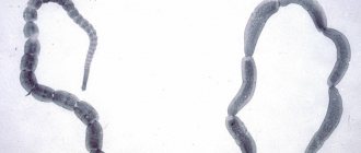
Features of tapeworms
The long body of worms belonging to this species consists of several segments that have their own reproductive organs, which increases the chances of survival when changing hosts. At the end of the worm there is a head with attachment devices.
Occasionally, cestodes with an undivided body are found. The intermediate host of parasites can be fleas, fish, pigs, and other domestic animals. The cat's digestive tract is usually inhabited by adults.
Most often it is pumpkin tapeworm, broad tapeworm, echinococcus, spirometra.
A pet becomes infected by eating raw meat or fish. Having attached themselves to the intestinal mucosa, parasites suck out beneficial substances and microelements from the animal’s body.
Visually, you can detect worms in a cat in the feces, in the anal area in the form of segments that externally resemble cucumber seeds up to 8 mm in size.
Tapeworms have their own special structure
How does infection occur?
The main way tapeworms are transmitted is through fleas. The larvae of these insects feed on helminth eggs, which are stored inside when the flea reaches the adult stage.
By licking its fur, the cat inadvertently swallows blood-sucking parasites, which leads to infection.
From the digested insect, the tapeworm ends up in the animal's intestine, where it attaches itself to the wall and begins its parasitic activity.
Symptoms and diagnosis
The presence of parasites in a cat is detected by many signs. As a result of irritation that occurs as the worms mature, the pet's behavior changes. Eggs and segments of the parasite come out with feces. The cat's condition is noticeably deteriorating. She begins to lick her genitals frequently. Cough and pneumonia may develop.
There is difficulty urinating, blood in the stool. Worms, feeding on healthy tissues of internal organs, cause dysfunction of the digestive system, exhaustion of the body, loss of appetite and weight loss. An additional negative effect is caused by toxic substances released during the life of the parasite.
Symptoms of helminthic infestation in a cat are in many ways similar to signs of other diseases. Therefore, you should not rush with deworming. If you suspect a parasitic infection, you should contact a veterinarian, who, with the help of special tests, can diagnose the disease.
Laboratory stool analysis does not always detect helminths. With a recent infection, young tapeworms are not yet capable of releasing eggs. The doctor may order an x-ray to identify parasites or a blood test to determine the presence of allergic reactions or anemia.
When infected, the cat loses its former activity.
An examination is carried out to detect other infectious diseases. Effective treatment is selected only after the type of worms parasitizing the cat’s body becomes known.
Treatment
It is important to diagnose helminthic infestation in time. Timely treatment will allow you to quickly get rid of parasites. Today there is a large selection of anthelmintic drugs for cats.
They allow you to easily cope with helminths at any stage of development, without causing a toxic effect on the pet’s body. Available in a variety of forms.
Your veterinarian will help you choose the right medicine and determine the dosage, taking into account the type of worms and the general condition of your pet.
Pills
There are drugs in tablet form that are used once. You can give the medicine forcibly by placing the tablet on the root of the tongue, or crush it and mix it with water and pour it into the mouth using a syringe. During the treatment process, it is important to ensure that the pet does not spit out the medicine. The most popular medications to help control tapeworms in cats are:
Kanikvantel fights all types of worms
- Holiday plus. The active ingredients are fenbendazole and praziquantel. By interacting with each other, they block the functioning of the muscle and nervous systems of the parasites, which causes the death of the latter. The remains of worms are excreted in the feces. The dosage depends on the weight of the pet - one tablet is designed for 10 kg of weight. If the cat weighs 5 kg, then he needs to be given half the tablet. Kanikvantel is suitable both for the treatment of helminthic infestation and for prevention. In severe cases of the disease, it is recommended to take the medication again after 2 weeks. The drug has a gentle effect. Even a significant excess of the dosage does not have a negative effect on the body. In rare cases, adverse reactions such as vomiting and diarrhea are possible. These symptoms usually go away on their own after a few hours. This product is contraindicated in pregnant cats and kittens under three weeks of age.
Drontal belongs to the group of the most effective drugs
- Drontal. Available in the form of white coated tablets. The active substances pyrantel embonate and praziquantel have a paralyzing effect on helminths, which leads to their death. Quickly absorbed into the intestinal villi, they penetrate all organs. Excreted through the kidneys. Easily tolerated by all cats, regardless of breed and age.
Pratel is effective against many types of parasites
- Pratel. Praziquantel and pyrantel are the main components. The drug is a yellowish anthelmintic tablet that is effective against many types of parasites, including tapeworms. By paralyzing the muscles and blocking the transmission of nerve impulses, it destroys adult helminths, as well as larvae.
Medicine for worms must be given exactly according to the indicated dosages.
Drops on the withers
This form of anthelmintic drugs is convenient when the animal refuses to take the medicine orally. Drops are applied to the occipital region once. Effective:
Profender is prescribed against many parasitic diseases
- Profender. An anthelmintic drug used for therapeutic and prophylactic purposes. Available in the form of a solution containing emodepside and praziquantel, components known to be effective against tapeworms. Having penetrated the animal’s skin into the body, the drug paralyzes the neuromuscular system of the parasite, causing its death. It can be maintained for a long time in therapeutic concentrations and is eliminated within 30 days.
Stronghold is capable of destroying adults and larvae
- Stronghold. A colorless solution containing selamectin as an active substance. Effective against many parasites, providing reliable protection for a month. If the specified dosage is observed, the drug is completely safe for the pet’s health.
Prazicide is a drug for complex use
- Prazicide-complex. Broad spectrum drops. The composition includes a complex of active substances - praziquantel, ivermectin, levamisole, thiamethoxam. Refers to moderately dangerous drugs.
Drops are applied to the occipital region once. Having parted the animal's fur, squeeze the contents of the pipette onto the skin in accordance with the recommended dosage. Manipulation should be carried out carefully so that the medicine does not get on your hands.
Suspensions
The medicine has a sweetish taste. Injected into the oral cavity using a syringe. The following drugs have found widespread use:
- Prazicide plus. Suitable for adult animals and kittens over 3 weeks old.
Dioctophimosis in carnivores
Dioctophimosis is a disease of carnivorous animals (mainly dogs), which is caused by round helminths that parasitize the renal pelvis, abdominal cavity, ureters and bladder.
Less commonly, they can be found in the liver, blood vessels and heart.
Infection of an animal's genitourinary tract by large nematodes leads to severe impairment of kidney function.
Helminths can reach a length of up to 40 cm. However, their width is no more than 7 mm. Their color varies from bright pink to red. The eggs of parasites are oval in shape.
Dogs are the definitive (main) hosts of parasites, and oligochaete worms act as intermediate hosts. Dogs and other sick animals excrete eggs in their urine, in which the larvae develop to the invasive stage. Dogs most often become infected by eating worms. From the intestines of dogs, the larvae travel through the blood to their localization sites.
Largely due to their large size, helminths in places where they are localized in the animal’s body have a significant mechanical, allergic and toxic effect. In especially severe cases, the affected kidney can completely atrophy.
With dioctophimosis, the urine of a sick animal becomes bloody or with clots of pus. Its release occurs in separate portions. These symptoms can lead to peritonitis.
During diagnosis, capillariasis may be detected.
No specific therapy has been developed to treat this nematode-associated disease. In severe chronic cases of the disease, surgical removal of helminths is indicated.
For subcutaneous injections, ivermectin is indicated at a dosage of 0.2 mg/kg once a day. The course of treatment is 5-7 days.
Types of helminths
The causative agents of helminthiases in cats are represented by two types: roundworms – Nemathelminthes
(class
Nematoda ), cause nematoses; flatworms - Plathelminthes
(class of tapeworms
Cestoidea and flukes Trematoda ), cause cestodiasis and trematodiasis, respectively.
Depending on the biology of helminths and the ways of their spread, 3 main groups are distinguished: geohelminthiasis, contact (contagious) and biohelminthiasis. Geohelminthiases are invasions whose pathogens develop without an intermediate host. The geohelminth eggs or larvae released from the body develop to an invasive stage in the soil.
Contact (contagious) helminth infections are transmitted directly from a sick animal to a healthy one, since the eggs enter the external environment already mature and do not need an additional development cycle.
Biohelminthiases are invasions in which the biological development cycle of the helminth necessarily takes place in the body of other living beings.
There are final ( definitive)
) hosts, in whose body the development of helminths occurs to the sexually mature stage, and
intermediate
- where the parasite is in the larval stage or reproduces asexually.
A paratenic host
is a potential intermediate organism in which development does not occur.
Transport
and
reservoir
are also distinguished .
Capillariasis in dogs
Capillariasis is a disease caused by round helminths that parasitize the cavity of the bladder. In this organ they attach to the mucous membrane and cause catarrhal inflammation.
This disease mainly affects dogs. The causative agent of the disease is a nematode with a thread-like body shape.
The development of parasites is characterized by the presence of intermediate hosts (earthworms). Earthworms eat the soil along with helminth eggs, from which larvae subsequently hatch in the intestines. Dogs become infected by accidentally eating worms. In this case, the worms are digested, and the parasite larvae are transferred through the bloodstream to the bladder. The disease tends to spread quickly, as dogs often mark their territory with urine, and each time a significant number of eggs are released.
Sick dogs experience frequent urination. There is often mucus or pus in the urine.
When diagnosing this disease, it is necessary not only to take into account the symptoms, but also to conduct a laboratory test of urine.
To do this, you need to take 100 ml of urine from the last portion, dilute it in a ratio of 1:1 or 1:2 with tap water, and leave it in a conical glass for 15-20 minutes. Then carefully drain the top layer and examine the sediment for eggs.
To treat capillariasis, a 10% aqueous solution of levamisole is prescribed, which should be administered intramuscularly to dogs at a dosage of 5 mg/kg for 2 days.
Also, for the treatment of this nematode in dogs, a 1% solution of ivermectin in the form of subcutaneous injections at a dosage of 0.02 ml/kg is indicated.
Fenbendazole is given to sick dogs along with food at a dosage of 20 mg/kg once a day. The course of treatment is 5 days.
What are worms?
Answering this question, it would be more correct to say: “not what they are,” but “who are worms”!
Worms (helminths in cats) are endoparasites, worms that live and reproduce in the body. These include nematodes (the most common in cats), tapeworms and flukes. Infection in cats occurs through the eggs of worms or larvae.
Cats have their own species-specific parasites. There is a group of especially dangerous helminths that pose a threat to human health.
What do worms look like in cats?
What do worms look like in kittens and cats?
Helminthiases (diseases caused by worms in cats) are very common. This situation is due to insufficient information among owners about the importance of the problem and, perhaps most importantly, to the large species diversity of cat worms.
Modern parasitology distinguishes 3 types of endoparasites:
- Flukes
(trematodes) are a dangerous, but not very common group of worms in cats. The worms are small in size, rarely exceeding 5 cm in length. Their body shape resembles a willow leaf. On the ventral side there is one or several suckers and in the depth of the sucker there is a mouth. During the development cycle, flukes have permanent hosts (most often fish and shellfish) and intermediate hosts (cats, dogs, other animals, humans). - Tapeworms
(cestodes) - they are very well adapted. Tapeworms in cats are long and flat. The body is in the form of a ribbon consisting of segments (cells) fastened together. Their length can be more than 3 meters! At one end there is a “head” with special suction cups and hooks that allow it to be securely attached to the intestinal mucosa. To reproduce the cestode in cats, the segment is separated from the tail end, releasing it into the environment with feces. Another animal or person swallows this member and also becomes infected with the parasite. - Roundworms
(nematodes) are the most common group of cat helminths. The size of roundworms in cats can vary, from a few millimeters to a meter. Outwardly, they look like a small earthworm, but this type of worm in cats is white or beige.
Coccidiosis in sheep
Coccidiosis is a disease caused by the activity of parasites that live in the internal organs of animals. Sheep are mainly affected by this disease.
The main symptom of the disease is diarrhea mixed with blood. In sheep, the disease can occur in acute and chronic forms and is accompanied by loss of appetite, indigestion, and fever. Sheep often lose weight, and lambs may even die.
To treat this nematode, add sulfamethacin or a 2% solution of sulfamidine to drinking water at a dosage of 1 g/l of water. The course of treatment is 7 days.
Measures to prevent this disease include maintaining cleanliness and quality feeding. It is necessary to periodically give young animals for prophylaxis along with food phenothiazine at a dosage of 0.5 g/kg 1 time per day for 3-5 days.
The main signs of nematodes are shown in these photos:
Onchocerciasis in cattle and horses
Onchocerciasis is a helminthiasis from the group of filariases (caused by nematodes), characterized by the formation of subcutaneous nodes, damage to the skin and eyes. In this disease, sexually mature onchocerci parasitize in the tendons and ligaments of animals, and the larvae (microonchocerci) are localized in the skin. The disease is widespread. It is mainly affected by cattle and horses.
Females of sexually mature onchocerci secrete a large number of larvae, which pass into the skin. Further development of the larvae takes place in the body of intermediate hosts (blood-sucking insects). The development of parasites in the definitive host occurs within 7-8 months.
Infection of animals occurs only near the breeding sites of midges, since infected female insects are unable to fly long distances.
Most often, this disease has a chronic form. There are no visible signs of this nematode-caused disease in cattle. In horses, the main symptoms of onchocerciasis are abscesses, cellulitis and lameness.
To make a diagnosis, it is necessary to conduct examinations of skin samples taken from the navel area. A sample 1.5-2 mm thick must be cut into small pieces and placed on a glass slide, then filled with saline solution. After 10-15 minutes, the pieces should be removed, and the preparation should be examined under a microscope to detect microonchocerci.
No special therapy has been developed for the treatment of this disease in cattle. In horses, worms are removed through surgery.
To prevent animals from becoming infected with onchocerciasis, it is necessary to protect them from attacks by invasive midges.
To combat blood-sucking midges, it is recommended to treat pets with pyrethroids, proteid or neocidol. Places where midges accumulate are also subject to treatment. For this, it is best to use byteks or difos.
Probability of human infection
New medications and therapeutic techniques are a 100% guarantee of success with timely treatment and the correct approach to diagnosis. The prognosis is favorable. In most cases, fairly reliable protection of a pet from infection with cestodes is the fight against blood-sucking ectoparasites.
Is there a chance of human infection? Yes, there are such cases, and this applies not only to echinococci and alveococci. Doctors have documented situations of human infection even with the cucumber tapeworm, which is relatively “safe” for people. After all, even inveterate neat freaks have a chance to swallow an infected flea. What can we say about small children who put any object of interest into their mouths! Thus, when caring for your sick pet, you need to strictly observe at least basic hygiene principles! If you have elderly people and/or small children at home, it is better to treat your pet on an outpatient basis. In cases where this is not possible, the animal must be completely isolated for the entire period of treatment.
By the way, getting a little off topic. Many parents assume that pinworms in their offspring are the result of contact with cats. Is this really true? No, this is a common misconception. Pinworms are specific to humans ; they do not parasitize cats at all. However, this does not in any way eliminate the need to constantly remind the younger generation of the importance of regular hand washing. This simple measure can save you from many problems.
Parascariasis in horses
Parascariasis is a helminthiasis in which parascaridae are localized in the small intestine, and sometimes in the stomach and bile ducts of the animal's liver. Parascariasis is observed everywhere. This disease mainly affects horses.
Parascarid eggs in the external environment reach the invasive stage in 1-2 weeks. From the moment a horse ingests parasite eggs until their sexually mature stage, approximately 1.5-2 months pass.
Equine parascariasis is more common in places with a humid climate and marshy soil. Mostly young individuals in stable and mixed housing are susceptible to the disease. Adult horses get sick less often.
Infection of horses with parascaridae is more often recorded in the autumn-winter period, after which a decrease in the disease is noted.
Horses are infected through the nutritional route by ingesting invasive parasite eggs in feed or water. Foals can become infected with parascariasis while nursing mares.
The symptoms of this disease, caused by nematodes, depend on the intensity of the damage, the age of the animals, the resistance of their body and possible allergic reactions.
The symptoms are most pronounced in foals, while in adults parascariasis is mainly subclinical. When parascarid larvae migrate through the blood in the first days after infection, enteritis and diarrhea are observed, then bronchopneumonia develops, accompanied by a cough, a short-term increase in body temperature and mucous discharge from the nose. In the presence of parascaridae in the intestines, digestive upset occurs, animals quickly lose weight and get tired quickly. The mucous membranes become pale, the composition of the blood changes, the number of red blood cells and hemoglobin decreases. Leukocytosis, eosinophilia, and accelerated erythrocyte sedimentation reaction are often observed. Sometimes foals experience convulsions or paresis.
To diagnose this disease, it is necessary to examine horse feces using the Fulleborn method. In foals infected with parascariasis, parascarid eggs cannot always be detected, so diagnostic deworming is recommended.
Piperazine, carbon disulfide and carbon tetrachloride are used to treat parascariasis.
Piperazine is indicated in the form of piperazine sulfate, piperazine hexahydrate in a dosage of 8-25 g per head (depending on the age of the animal) twice for 2 consecutive days with food or water.
Carbon tetrachloride should be administered in a dosage that depends on the age of the animal. For foals from 3 to 7 months, this drug is indicated in a dosage of 5-10 ml, from 7 to 12 months - 10-15 ml, from one year to 2 years - 15-20 ml, for adult horses - 25-40 ml. In case of significant helminth infestation, it is recommended to give a saline laxative after carbon tetrachloride. Before administering the drug, animals are given a 16-18-hour fasting diet. During the deworming period, concentrates should be completely excluded from the diet. Manure must be removed daily and disinfected using the biothermal method. Approximately 10 days after deworming, the stable should be treated with a hot 5% carbolic acid solution.
For prevention, scheduled deworming of horses should be carried out in the fall and spring. It is prohibited to feed roughage to horses from the floor. Animals can be given water only with clean drinking water.
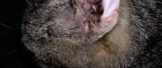
HOOKYLOSTOMIS
Ancylostoma tubaeforma is a hookworm, a small round helmin of the Chromadorea class, 1-2 cm long, which has two chitinous plates with three hook-shaped teeth on the oral capsule for fixation in the intestine.
Routes of infection
- Ingestion of larvae with food (feed).
- Active penetration of larvae through the skin.
- Hunting for rodents, in which the larvae can remain viable for up to 10 months.
Life cycle
Hookworm is a geohelminth
.
The eggs hatch into larvae after 10 days in the soil. If the larva enters the cat’s digestive tract with food, it quickly develops into an adult, sexually mature individual. When introduced through the skin, the larva travels a long way through the circulatory system, reaching the respiratory system and bronchi, rises up to the pharynx and is swallowed by the animal into the stomach. From them, adult individuals develop, which are attached by hooks to the intestinal wall. The hookworm is a hematotroph
- it feeds on blood. One infected cat can harbor up to several hundred hookworms. The females of the parasite lay a huge number of eggs every day, which fall into the soil with the animal’s feces.
The definitive host for Ancylostoma tubaeforma is the cat.
The paratenic host for Ancylostoma tubaeforma is rodents
Humans are not infected with this type of hookworm. Ancylostoma duodenale is pathogenic for humans.
Symptoms
The most dangerous symptom of hookworm infection is chronic blood loss with the development of anemia
. The sites of helminth penetration in the small intestine constantly bleed. With mass infestation, feces may become black (melena), and there may be profuse bleeding that threatens the life of the animal. A blood test shows a decrease in hemoglobin, the number of red blood cells and an increase in reticulocytes, as the red bone marrow tries to compensate for blood loss. In addition, the attachment of the parasite to the intestinal wall causes inflammation, which is manifested by enteritis, indigestion, diarrhea, the animal loses weight, protein-mineral metabolism is disrupted, and general health and coat condition deteriorate.
Parafilariasis in horses
Parafilariasis is a disease of horses in which helminths parasitize the subcutaneous tissue of animals. The causative agents are white thread nematodes.
The course of this disease is characterized by seasonality. Basically, the first cases of infection occur in April, and the peak incidence occurs in July-August. The higher the air temperature, the more pronounced the disease manifests itself. The largest number of horses become infected as a result of prolonged stay on pastures. Most often adults become ill.
Helminths, moving in the subcutaneous tissue, actively drill into the skin of the animal. In places of lesions, tubercles form on the skin of infected horses. Over a few days they gradually increase to the size of a pea. They appear mainly on the withers and shoulder blades, in the back and ribs, on the neck, and sometimes on the lower back and croup. Then, in these places, in sunny weather, holes and wounds form, through which blood leaks from broken capillaries and small vessels. At night the bleeding stops. After some time, the wounds heal and the tubercles disappear.
Bleeding is not observed daily throughout the summer season, but intermittently at different times. There can be more than 150 bleeding wounds on a horse's body. With such intensity of helminth damage, the horse loses a large amount of blood. When a collar or saddle comes into contact with wounds on the horse's skin, phlegmon appears.
In places where parasites live, abscesses that arise as a result of inflammation of the subcutaneous tissue can form without external influence.
To establish a diagnosis, it is necessary to take into account the seasonal characteristics of the invasion, as well as the symptoms of this type of nematode. It is recommended to conduct a blood test from emerging wounds in order to detect eggs and larvae of nematodes. To do this, a drop of blood on a glass slide should be diluted with 10 drops of distilled water and, without covering it with a coverslip, placed under a microscope. Blood can be drawn into a centrifuge tube and diluted with water in a ratio of 1:10, and the resulting sediment can be placed under a microscope. There are no microfilariae in the circulating blood, so there is no need to distinguish them from microsetaria.
To treat this disease, it is recommended to inject a weak aqueous solution of potassium permanganate subcutaneously into the affected areas in a ratio of 1:1000.
Ivomec should be administered subcutaneously at a dosage of 1 ml/50 kg or ivermectin at a dosage of 0.2 mg/kg.
To prevent parafilariasis, it is recommended to use various insecticides.
How to rid cats of worms
Contact your veterinarian. Drugs for the treatment of helminthic infections in pets are available in different forms. Most often, tablets and drops are purchased. They are less toxic and easy to use.
The most popular are:
- Advocate. Effective drops that need to be dripped onto your pet's withers. It will help get rid of not only external, but also internal parasites. Acts against nematodes. Can be used once. Apply to dry skin.
- Helmimax. The tablets are effective against cestodes and nematodes. Can also be used for mixed invasions.
- Helminthal. The form of the drug is drops. Apply to the withers. Fights tapeworms and roundworms.
- Dirofen. This is a single use tablet. The dosage is calculated for an animal weighing up to 5 kg. It is recommended to treat helminthiasis twice, with a break of 10 days.
- Drontal. The tablets are effective against tapeworms and roundworms.
- Inspector. Effective drops that work against most types of parasites. Apply to dry and clean skin of the cat, in a place where she cannot lick the drug. After application, do not bathe the animal.
- Milbemax. They work against nematodes and cestodes. .
- Prazicide. The tablets have a wide spectrum of action.
- Profender. Broad spectrum drops. Apply to the pet's withers. The skin should be clean and dry. They are used for treatment and prevention.
- Stronghold. Effective drops on the cat's withers. They work against all types of parasites. At the same time, they will destroy fleas. It can be used not only for treatment, but also for prevention. They work for a month. If necessary, repeat treatment.
Sarcosporidiosis in dogs and cats
Sarcosporidiosis is a protozoal disease of dogs and cats caused by various species of Sarcosporidium, which are localized in the mucous membrane of the small intestine of the definitive (primary) hosts. A person can also become infected.
In intermediate hosts, parasites are mainly found in the muscles and heart. In this case, the disease proceeds subclinically, since the cystic stages of the parasite (sarcocysts) do not cause pathological changes.
The process of parasite reproduction in dogs and cats occurs in the epithelial cells of the mucous membrane of the small intestine, resulting in impaired digestive function.
Sarcocysts, parasitizing dogs, can cause severe sarcocystosis in intermediate hosts (cattle, sheep, pigs).
Sarcosporidiosis of carnivorous animals is widespread, but these diseases are diagnosed in practice quite rarely, since there are no clinical signs characteristic only of these diseases.
Sarcocysts develop with the obligatory participation of two hosts - the final and intermediate.
Animals are susceptible to this disease regardless of age.
The severity of the disease depends on the number of sarcocysts in the animal’s body.
In case of mass infection, damage to the intestinal mucosa, impaired digestion and absorption of nutrients are observed. In such cases, the animals become depressed and experience decreased appetite and diarrhea for several days.
To diagnose this disease, it is necessary to take into account epidemiological data, symptoms and results of scatological studies. To conduct scatological studies, fresh stool samples should be used, in which Sarcosporidium sporocysts are often found in sporulated form.
To treat this disease caused by nematodes in dogs and cats, sulfonamide drugs should be used at a dosage of 60-100 mg/kg once a day. The course of treatment is 5 days.
A good therapeutic effect is achieved by using the chemical coccide at a dosage of 24 mg/kg once a day. The course of treatment is 5 days.
Coccidiovit is indicated in a dosage of 150 mg/kg 2 times a day. The course of treatment is 3-5 days.
To avoid contracting sarcosporidiosis in cats and dogs, you should avoid feeding them raw meat. It is recommended that dogs be regularly screened for the presence of sporocysts.
Diagnosis of helminthiasis
To find out which tablets to give your dog against worms, and to determine the degree and severity of the infection, you need to do the following:
- Fecal analysis, which shows whether dogs have worms and their type. Moreover, if worm eggs in dogs are detected quickly, it takes time to find out which worm has infected the animal.
- Blood analysis. Plasma testing determines antibodies, inflammatory processes and other pathologies that develop during helminthic infestation. With its help, the doctor finds out the extent of the damage, which helps prescribe treatment.
- Chest X-ray, ultrasound of the heart - the veterinarian prescribes if pulmonary or cardiac infection is suspected. X-ray and ultrasound will show the presence/extent of the lesion.
Setaria in farm animals
Setariasis is a helminthiasis that is caused by nematodes that parasitize in the abdominal cavity (mature individuals), in the brain and spinal cord, in the eyes (juvenile forms) and in the blood (microsetaria).
Many types of farm animals are susceptible to this disease, including cattle, sheep, goats, and horses.
Setaria develop with the participation of intermediate hosts (mosquitoes and flies). The development of the parasite in the definitive host occurs within 6 months. Juvenile forms of helminths parasitize in the brain and spinal cord of sheep, and its mature forms are localized in the abdominal cavity of cattle.
Infection occurs by inoculation of infective larvae by blood-sucking insects on pasture. The greatest number of infections occurs during the flight of insects.
The main symptoms of this nematode, caused by setaria nematodes, depend on the degree of damage. In horses and cattle, clinical manifestations have not been studied. Paresis and paralysis of the limbs are observed in sheep. When the brain is damaged in sheep, movement coordination disorder occurs, which often leads to the death of the animals.
To diagnose setaria in horses and cattle, it is necessary to conduct a blood test to detect microsetaria.
No special therapy has been developed. Control and prevention measures include preventing contact between definitive and intermediate hosts. It is important to combat insects that carry setaria.
Routes of infection
Parasitic eggs are always found in the environment. These are vegetables, water, fish, grass, dust, meat. Most often, a pet becomes infected from homeless people.
Main routes of infection:
- Raw water. Worm eggs live in damp, often river water.
- Mother's milk. Infection of kittens with helminths occurs during the first feeding (provided that the mother is infected).
- Intermediate hosts. They can be fleas, fish, rodents, crustaceans.
- Feces. When cats wash themselves, they often lick feces from their paws. This is how infection occurs.
- Grass, soil. Helminth eggs are found on the grass and in the soil. As the animal moves, they stick to its paws.
The routes of infection by parasitic worms in a cat will directly depend on the type of worms.
Spirocercosis in dogs
Spirocerciasis is a disease of dogs caused by roundworms that parasitize in inflammatory tubercles of the esophagus, stomach, aorta and lungs.
Beetles act as intermediate hosts and eat eggs released by sick animals. It is in the body of the beetles that the parasite larvae reach the invasive stage.
Dogs become infected by eating infested beetles.
There are reservoir hosts (birds, reptiles, etc.) that feed on beetles. The larvae remain in their body for a long time and serve as a source of infection for dogs. In the dog's body, the larvae travel with the blood to the localization sites, around which swellings appear with a hole at the top, from where the eggs of mature helminths come out.
Helminths reach sexual maturity in about 5 months.
The rapid spread of the disease is due to the presence of a wide range of reservoir hosts.
The presence of swelling in the aorta, esophagus, stomach and other vital organs significantly impairs the properties of these organs. Symptoms of the disease reflect the pathology of those organs in which the parasites are localized, therefore, disturbances in the rhythm of blood flow, swallowing, coughing, etc. are observed. Diagnosis of this disease requires a set of studies, including laboratory ones. Fecal examination is carried out using the Fulleborn method. To detect swelling, fluoroscopy of organs can be performed.
No specific therapy has been developed, but levamisole and ivermectin can be used for treatment. Animals are injected intramuscularly with a 10% aqueous solution of levamisole at a dosage of 5 mg/kg for 2 days. A 1% solution of ivermectin is indicated as a subcutaneous injection at a dosage of 0.02 ml/kg. The course of treatment is 3-5 days.
To prevent spirocerciasis in dogs, animal feces should be regularly disposed of by burning or burying.
Tapeworms (worms) in cats: symptoms, treatment, routes of infection
Even domestic cats can sooner or later become victims of worms, and tapeworms are one of the common intestinal parasites. There are several types of worms in cats.
The most common are worms, which are transmitted by ingesting fleas, but there are also varieties that infect cats that feed on wild animals (birds, rodents and lizards).
How can a cat become infected with tapeworms?
The most common way cats become infected is by ingesting infected fleas while licking their fur. Cats ingest about 50 percent of the fleas that may be present on their fur when they groom themselves. Once a cat swallows an infected flea, the tapeworm larvae enter the cat's intestines, where they develop into adults.
Outdoor cats that hunt birds, rodents, or lizards are also at risk of contracting tapeworms. If a cat ingests animals infected with tapeworms, she may also become infected.
What are the signs and symptoms of heartworms (worms) in a cat?
An adult tapeworm is a long, flatworm that can live in a cat's intestines for months. Small segments (segments) filled with eggs are detached and passed out in her feces. Although these segments are no longer living, they remain mobile for some time.
You may not know if your cat has tapeworms because in most cases there are no or subtle symptoms. However, you can spot the following symptoms of worm infestation:
- You may see parts of the tapeworm under your cat's tail or in her feces.
- Itching that causes the cat to repeatedly lick or bite the area under the tail, or "roll" on the ground in an attempt to relieve the itching
- Weight loss
- General malaise
A drug such as Profender® against worms for cats is a simple and easy way to fight worms. Profender® acts on the most common types of worms that are commonly found in cats. Profender® is applied to the back of the cat's neck (withers) to prevent the cat from licking the drug.
If you think that deworming tablets are a simpler solution, then Drontal® for cats is also registered for a wide range of the most common cat worms and is available in one dosage - one tablet per 4 kg of cat weight.
Maintaining a regular worming schedule will help protect your pet from these uninvited guests. Regular preventive maintenance is recommended for the health and protection of the cat.
Toxascariasis in dogs and cats
Toxascariasis is a disease caused by a nematode that parasitizes the small intestine and sometimes the stomach of animals.
This disease mainly affects adult dogs and puppies over 6 months of age, but it also occurs in cats.
The nematode is light yellow in color and spindle-shaped. The development of the parasite occurs in a direct way. In the eggs released along with feces into the external environment, larvae develop, as a result of which the eggs become invasive.
After entering the dog’s digestive tract, the larvae of such eggs are released from the shell, localized in the epithelial wall of the intestine and continue to develop there.
After some time, the parasite larvae move into the intestinal lumen, where after about 3-4 weeks they reach sexual maturity.
Nematodes have mechanical and toxic effects on the body of dogs and cats. With severe damage in animals, blockage of the intestinal lumen may occur, and sometimes rupture of its walls, followed by peritonitis.
Sexually mature nematodes often enter the bile duct and bile ducts of the liver, pancreatic duct, and stomach from the intestine. As a result, the functional activity of these organs is disrupted.
Sick dogs experience severe emaciation, mucous membranes become pale, and indigestion often occurs.
The animals completely lose their appetite. Diarrhea may alternate with constipation, and vomiting is sometimes observed. Abortions often occur in pregnant bitches.
To diagnose this nematode in cats and dogs, it is recommended to use the Fulleborn method.
Piperazine and tetrachlorethylene are used to treat toxascariasis.
Piperazine is indicated at a dosage of 0.2 g/kg for 2 days. This drug should be given to animals mixed with food in the morning and evening.
Tetrachlorethylene is prescribed in gelatin capsules at a dosage of 0.1-0.2 ml/kg. The course of treatment is 3 days.
3 days before deworming, fats should be completely eliminated from the animal’s diet.
TOXOCAROSIS
Toxocara cati (Toxocara mystax) is a “cat” toxocara, a helminth of the Nematoda class. Size from 3 to 10 cm, females are larger than males. A distinctive feature of the male is the curved rear end. In the area of the head, a sexually mature individual has wings of the cervical type, the mouth opening is surrounded by three lips. The eggs are round in shape, brown in color with a thick, rough shell.
Routes of infection
Toxocara eggs are ubiquitous.
- An animal on a street walk can become infected by licking city/village dust from its fur or paws (oral-fecal route of infection).
- Mousecatching cats are at risk because Toxocara uses rodents as possible intermediate hosts.
- When eating raw meat in a diet contaminated with helminth eggs.
- A person can bring eggs into an apartment on shoes, clothes, or bags.
- When feeding kittens, as the medications can pass into mother's milk. Unlike canine Toxocara, feline Toxocara does not penetrate the placental barrier and does not infect kittens in utero.
Life cycle
A cat swallows Toxocara eggs directly by licking or eating paratenic (transport) hosts - mice, rats. The egg hatches into a larva, which bores through the intestinal wall and enters the bloodstream. With the bloodstream they are carried throughout the animal’s body, mainly settling in the lungs. Another part of the larvae reaches other organs (liver, lymph nodes, spleen, brain), where they are encapsulated and do not develop. In the lungs, Toxocara larvae rupture the alveoli, penetrate the bronchioles and move up to the larynx. During this period, the cat may cough and swallow the larvae along with saliva back into the digestive tract, where they develop into mature adults that produce eggs. The cat begins to release Toxocara eggs into the external environment with feces. Encapsulated larvae can periodically “wake up” and migrate again with the bloodstream to other organs. In addition, during lactation they pass into breast milk and within 3 weeks mature worms develop in the kittens’ intestines.
The definitive (final) host for Toxocara cati is the cat.
The paratenic host for Toxocara cati is rodents, worms, cockroaches, birds, humans, in which Toxocara cati does not develop into a sexually mature individual, remaining in the form of an encapsulated larva for up to 10 years.
Symptoms
Symptoms are nonspecific. Cats may experience decreased activity and loss of appetite. Periodic digestive disorders and enlarged lymph nodes are possible. Animals develop anemia and their immunity decreases. Toxocariasis causes disruption of mineral and protein metabolism. The condition of the coat deteriorates and dandruff appears. Massive infestation by Toxocara is especially dangerous, since worms, clogging the intestines, can cause obstruction, including intestinal rupture, the development of peritonitis and the death of the animal.
Tominxosis in dogs and cats
Tominxosis is a disease of dogs and cats caused by filamentous roundworms that parasitize the bronchi, trachea and nasal cavity. In this case, helminths have not only a mechanical, but also an allergic effect.
Intermediate hosts (earthworms) also take part in the development of helminths. Approximately 2-3 weeks after being excreted in feces, Tominx eggs become invasive. Earthworms swallow them along with the soil. Then the larvae hatch from the eggs and settle in the body of the earthworms.
Dogs and cats become infected when they accidentally eat infected worms. Helminths reach sexual maturity approximately a month after entering the body of the definitive host, and the life expectancy of these parasites is 9-10 months.
Bronchopneumonia is often observed in sick animals, and when a secondary infection occurs, purulent bronchopneumonia and abscesses in the lungs develop.
Breathing becomes difficult, wheezing, coughing and mucous discharge from the nostrils appear.
This disease is especially difficult for young individuals.
To diagnose the disease, feces are examined using the Fulleborn method to detect parasite eggs.
Levamisole, ivermectin and nilverm are used to treat dogs and cats.
A 10% solution of levamisole is prescribed at a dosage of 5 mg/kg once a day. The course of treatment is 2 days.
A 1% solution of ivermectin is administered subcutaneously at a dosage of 0.02 ml/kg once a day. The course of treatment is 2-3 days.
Nilverm is prescribed at a dosage of 3-5 mg/kg 2 times a day. The course of treatment is 7 days. The drug must be given in mixture with food.
To avoid infecting animals with tominxosis, the possibility of dogs and cats eating earthworms should be excluded. Do not leave food in pets' dishes, as worms may crawl into them.
Worms in cats: ???? symptoms and treatment of helminths, drugs
Contents
It happens that a beloved pet receives a gift from nature of neighbors in the body - worms. Not the most pleasant neighborhood, from which the animal’s body will suffer for a long time if the owner does not undertake deworming. Worms in a cat are a serious disease that depresses all systems of the body: the animal loses weight, eats poorly, and becomes very tired.
Where does such a misfortune come from? Such unkind guests easily enter the body, but leave with reluctance. If you suspect unwanted neighbors, it is better to visit the veterinarian and find out who decided to destroy the cat from the inside.
Types of worms in cats and cats
Worms in cats come in different sizes and shapes, and they feed and reproduce differently.
They have long been classified into three categories: flukes, tapeworms, and roundworms. Let's study the enemy! Name Features Symptoms in a cat
| Lungworms | This infection enters the cat if it drinks from a dirty source or eats crustaceans. Tapeworms destroy the lungs with cyst-like formations |
|
| Cucumber tapeworm | The parasite can easily take up residence in both the cat’s body and the human body. In a cat, the size of a tapeworm rarely exceeds a length of 30 cm. The danger is expressed by damage to the intestinal walls. The parasite can be brought by fleas and lice eaters. | The pet is vomiting, losing weight, and is irritable. My stomach is constantly rumbling and I have diarrhea. |
| Wide tapeworm | The parasite grows in the body up to 1.5 meters in an animal, and up to 12 meters in a person. You can become infected by eating raw fish, crustaceans and drinking river water. |
|
| Liver fluke | The fluke has chosen the liver as its home and does not disdain the pancreas and gall bladder. To acquire such a neighbor, you can eat raw fish. | The vomit is colored in shades of yellow, there is no appetite, the animal is weakened. Diarrhea appears and the temperature may rise. Complex treatment is used here, due to the ineffectiveness of standard treatment. |
| Causative agents of alveococcosis | The parasite reaches a length of 4-5 mm and does not pose a danger to the cat. For many years this neighborhood has been quite peaceful, but as soon as a person becomes infected, trouble will strike. It is difficult to make a diagnosis; the patient faces death due to metastases to various organs, and a liver tumor develops. The worm can enter the body as a result of eating a rodent. | There are no symptoms. |
| Toxocars | The localization of five-centimeter helminths is the esophagus, gall bladder and liver. The source of infection is food, and cases of intrauterine infection are also known. The parasite is dangerous for kittens, as the rapid development of toxocara begins, which is fraught with ruptures in the small intestine. | No symptoms observed |
| Hookworms | The size of these guests of the cat’s body does not exceed 1 cm. Hookworms choose the intestines as their place of residence, and blood as their source of nutrition. Helminth eggs can enter the body with food, and the larvae can even penetrate the skin of a pet. | Appetite is reduced, the animal is lethargic and apathetic to everything. The stool is liquid and there is visible blood in it. The cat is vomiting and coughing a lot. |
These are the main parasitic creatures that can seriously complicate the life of a pet and its owner.
Is it possible to get worms from a cat?
There are approximately 32 known varieties of worms in cats , which will not deny themselves the pleasure of settling in the human body. The consequences of such proximity in animals and humans can manifest themselves differently. If a pet’s body can calmly coexist with some types of helminths, then in some cases the consequences for humans are serious.
Infection occurs after the eggs or larvae of worms enter the oral cavity. Basically, they get there through the hands. Even if several hours have passed after contact with the pet, parasite eggs remain on the hands.
A cat, licking its fur, spreads pathogens through it from its anus, where the larvae and eggs of helminths accumulate.
A person pets a furry friend, forgets to wash his hands with soap, and soon finds out that he has new little “friends” in himself.
Worms are transmitted not only from direct contact with the cat, but also through his things and litter box.
Worms dangerous to humans
Let's look at helminths that are transmitted from cats to humans and are dangerous:
- Flatworms cause a disease called opisthorchiasis. The parasite settles in the liver, gall bladder and pancreas, affecting the ducts of these organs. You can get sick by eating raw or poorly cooked fish, mainly from the carp family;
- The wide tapeworm of the cestodoses group causes diphyllobothriasis. The worm occupies the small intestine and grows up to 10 meters. Iron deficiency anemia and dyspeptic disorders gradually develop. Raw freshwater fish can also become a source of infection;
- Echinococcus in the larval stage can seriously affect the internal organs of a person. Cats also carry the dangerous disease alveococcosis to humans. Contact with a sick animal can be fatal for people.
Hygiene remains the main barrier to the transmission of such diseases.
Signs of worms in a cat
Worm infestations do not manifest themselves immediately: the development of symptoms is directly proportional to the amount of infection in the body.
Symptoms of worms in cats include a number of signs:
- depressed and apathetic state;
- problems with appetite;
- weight loss;
- fatigue;
- slow development in kittens;
- unusual condition of the coat, its disheveled appearance;
- noticeable yellowness of the mucous membranes;
- on palpation there is noticeable enlargement of the liver (trematodes);
- anemia of the mucous membranes (cestodia);
- constipation gives way to diarrhea, vomiting;
- cough;
- frequent licking of the anus due to itching;
- intoxication in the form of convulsions, paresis of the limbs;
- premature birth and sometimes miscarriage;
- blood in stool.
The list is quite large, symptoms do not appear individually, they are a whole set of signs.
Treatment of worms in cats
Anthelmintic treatment is prescribed by a veterinarian after testing. It is required to accurately identify the enemy and locate the source of the lesion.
Why is it necessary to consult a veterinarian? Because each anthelmintic medication acts on one type of parasite, and it is not so easy to remove worms from a cat But there are also complex drugs that act on a certain number of helminths.
The treatment regimen is simple: the drug for worms is given once in the morning, then the dose is repeated after 10 days. Deworming is also carried out 10 days before vaccination. Cats are dewormed 2-3 weeks before and 3 weeks after birth.
Remedies for worms in cats
If you suspect that your cat has worms, then you need to study the list of means that can be used to deworm the animal. These are effective medications in many cases.
Trichocephalosis in dogs
Trichocephalosis is a disease of dogs caused by round helminths that parasitize the mucosa of the cecum and large intestine. Trichocephalosis is widespread.
The causative agents of this disease are whipworms, which parasitize dogs.
Animals become infected by ingesting eggs in food and water. In the intestine, the eggs hatch into larvae, which then temporarily move into the intestinal mucosa and then return to the intestinal cavity. Helminths reach sexual maturity in 3 months and live for several months.
Nematodes feed on blood from the intestinal wall, which leads to serious pathological changes.
Sick animals become depressed and rapidly lose weight. Digestive dysfunction often occurs, with constipation or diarrhea. To diagnose this disease, feces are examined using the Fulleborn method.
Praziquantel, febantel, flubendazole, fenbendazole and mebendazole are used to treat trichuriasis.
Mebendazole should be given to animals with food at a dosage of 60-100 mg/kg once a day. The course of treatment is 3 days.
Praziquantel is administered once with food at a dosage of 5 mg/kg.
Febantel should be given to animals with food at a dosage of 0.01 g/kg once a day. The course of treatment is 3 days.
Fenbendazole is prescribed once in a mixture with food at a dosage of 25 mg/kg.
Flubendazole is given to animals at a dosage of 0.25/kg once a day. The course of treatment is 3 days.
To prevent dogs from becoming infected with trichuriasis, it is necessary to maintain hygiene in keeping and feeding animals, as well as promptly diagnose the disease and carry out deworming.
Diagnosis
Based on the above symptoms, tests are prescribed to identify parasites. To detect helminths, scatological studies are carried out. These tests help detect nematode eggs in animal feces. But if a cat or dog has recently become infected with helminths, then during the first test, parasite eggs may not be detected, due to the fact that there are still only a small number of them. In this case, it is better to conduct this study 3-4 times a month, which will give a more accurate result
Uncinariasis in dogs and cats
Uncinariasis is a helminthiasis in dogs, but cats are also sometimes infected. The disease is widespread.
The causative agent of this disease is a nematode that has a powerful oral capsule with two cutting blades. The hatching of larvae from eggs occurs in the external environment.
Cats and dogs become infected with nematodes through nutrition (with food) or through the active penetration of larvae through the skin, followed by their passage through the lungs into the intestines. It is in the intestines that parasite larvae reach sexual maturity.
Uncinaria significantly injure the intestinal walls of the animal, thereby causing capillary bleeding.
Mostly young individuals are susceptible to this disease.
Characteristic symptoms of uncinariasis are anemia, disturbances in the secretory and motor functions of the digestive organs, then bloody diarrhea and exhaustion of the animal can be observed. The disease lasts from 1 to 4 weeks and most often ends in death, but can also become chronic.
To make a diagnosis, it is necessary to conduct scatological studies to detect eggs or larvae of the parasite.
Carbon tetrachloride and tetrachlorethylene are used for treatment.
Carbon tetrachloride is prescribed in a dosage of 0.3 g/kg, but you should be especially careful, since toxic effects are possible as a result of exposure to this drug. The course of treatment is 2-3 days.
Tetrachlorethylene is indicated in a dosage of 0.1-0.2 g/kg 2 times a day. The course of treatment is 2 days.
These drugs should be given to animals along with food. For 3 days before and after the use of anthelmintic drugs, it is necessary to exclude fatty foods from the pet’s diet.
DIPILIDIOSIS
Dipylidium caninum - pumpkin, cucumber or dog tapeworm. This is a white or slightly yellowish cestode, 20-50 cm long, 3 mm wide. The scolex is equipped with four oval suckers and a club-shaped retractable proboscis with 4-8 transverse rows of hooks.
Routes of infection
- Ingestion of infected fleas, lice eaters.
Life cycle
Mature segments, breaking away from the strobila, actively emerge onto the perianal folds, where they are destroyed, and the eggs are dispersed into the external environment and onto the animal’s fur. Flea larvae
and
lice eaters
eat oncospheres (eggs), but no changes occur in them.
The oncosphere begins to develop into a cysticercoid in the pupa of an ectoparasite insect. An animal becomes infected by licking and gnawing fleas
,
lice eaters
, and swallowing them. They are digested, and the released cysticercoids attach to the wall of the small intestine, developing into adults.
Definitive owner
– dogs, cats, foxes, wolves, jackals and other carnivores.
Intermediate host
– ectoparasite insects (fleas, lice eaters, etc.)
Humans are the facultative host due to accidental infestation (flea ingestion).
Symptoms
Symptoms are nonspecific; more often the disease occurs latently. The general condition is depressed, motor activity and appetite decrease, the animal loses weight, and the quality of the coat deteriorates. With significant infestation, manifestations of enteritis (diarrhea, constipation, bloating) are possible due to the irritating effect of the tapeworm's suckers and hooks on the intestinal wall. Motile proglottids can be found in feces. With massive invasion, intestinal obstruction can form.
Habertiosis in sheep
Habertiosis is a helminthiasis of ruminants, mainly sheep, caused by nematodes that parasitize the large intestine. This disease occurs everywhere.
The nematodes that cause this disease are large in size, white in color, and have a characteristic blunt and beveled head end. The development of parasite larvae takes place in 5-7 days. Infection occurs through the nutritional route (by ingesting infective larvae).
After ingestion by an animal, the larvae develop in its body to mature helminths.
Most often, young individuals are susceptible to this disease.
Outbreaks of habertiosis occur in the spring (March-April), especially during rainy periods.
Sick animals experience disheveled hair loss, rapid weight loss, anemia, and diarrhea.
To make a diagnosis, infective larvae isolated from the feces of infected animals are grown.
For treatment, phenothiazine is prescribed at a dosage of 0.5 g/kg twice. The drug should be given every other day.
Nilverm is also prescribed at a dosage of 0.02 g/kg once.
For prevention, it is recommended to deworm young sheep before turning them out to pasture (January-February). You can give animals phenothiazine in a smaller dosage mixed with table salt.
Why are worms dangerous for cats?
A cat's health is at risk when there are high numbers of worms in the body. As we have already understood, the symptoms when cats are infected with worms are very non-specific. They can also be confused with bacterial, viral and non-communicable diseases. Regular prevention is important to prevent critical disorders.
• Can a cat, cat or kitten die from worms?
Worms in cats and kittens can cause death if the infestation is severe, but, as a rule, owners manage to notice the symptoms and cure the animal.
Chiostrongylosis in pigs
Chiostrongylosis is a helminthiasis of pigs caused by nematodes that parasitize the stomach. This disease is widespread everywhere. The emergence of larvae from eggs occurs in the external environment. They are able to remain viable in water for more than 8 months.
In the body of the host (pigs), the parasite larvae penetrate into the thickness of the gastric mucosa, where after 3 weeks they reach sexual maturity. Infection occurs through the nutritional route. Chiostrongylosis mainly affects adults. Meat breed pigs are least likely to become infected. The disease appears in the spring and reaches its peak in the fall.
Sick animals are lethargic, most of the time they lie buried in the bedding, their fur becomes disheveled. Pigs refuse food. Bloody diarrhea is common.
To treat this disease caused by nematodes, thiabendazole is prescribed once with food at a dosage of 100 mg/kg. Long-term use of this drug is possible in a 0.05% concentration as a feed additive. Approximately 10 days before slaughter, this drug should be excluded from the diet.
Treatment
How to treat your pet to get rid of worms once and for all? As a rule, treatment will be aimed at removing the parasites themselves from the body and relieving the symptoms accompanying the invasion. The main role here will be played, of course, by the use of anthelmintic drugs. Nowadays, veterinary pharmacies offer a wide range of drugs that help in the fight and removal of worms; they differ only in the basis of the active substance. It can be pyrantel, praziquantel or febendazole.
Most anthelmintics are sold in the form of tablets or suspensions that are easy to take. In addition, we should not forget that removing worms from a kitten requires fundamentally different means, so separate medications with a different dosage are produced for them. Remember that treatment of worms in cats at home will only be successful if it is started on time, and for this their presence must be quickly recognized.