Fistula is a life-threatening disease that is easy to identify when examining the animal. An open wound appears on the skin, forming a channel between the surface of the body and internal organs, anatomical cavities in the body. If you see an open wound with an unpleasant odor, do not look on the Internet for recommendations on how to treat a fistula in a cat; it is advisable to immediately show it to a veterinarian without delay. Some owners share successful experiences in treating the disease with antibacterial ointments or folk remedies. Such therapy may not help, but may worsen the animal’s condition and provoke the development of sepsis. Antibacterial ointments, applied without removing infected tissue, help with fistula-like abscesses. They can be distinguished upon examination by a specialist.
Signs of an abscess in a cat:
- A few days before the abscess appears, the tissues swell, become inflamed, and become warmer to the touch than areas of the body nearby.
- The wound has a regular round shape without branches.
- After the abscess matures, the tissues become soft and loose.
- Pus and some ichor are released.
- After a few days, it becomes noticeable that the wound is healing.
Signs of fistula:
- From above, the pathological hole looks like a branched funnel.
- The wound releases pus, ichor, possibly feces or urine.
- The hair around the wound falls out and the tissue becomes inflamed.
- An unpleasant odor is clearly felt.
- The wound does not heal.
If you have an abscess or fistula, you should not try to squeeze out the pus on your own, break through the abscess, or use anti-inflammatory or wound-healing folk remedies. This can lead to infection of internal organs and tissues around the wound. A veterinarian should help!
Where can a fistula appear in a cat?
Deep, pus-producing wounds with jagged edges occur in different parts of the body. Owners may mistake a fistula on a cat's paw or tail for a wound received in a fight with other animals. This is also a reason to take a trip to the veterinarian's office.
The disease can develop:
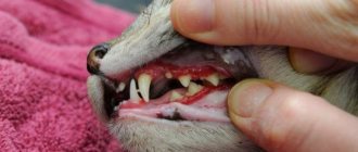
- On the cheeks, jaws - appears during inflammatory processes in the salivary glands, dental diseases. If detected early, it can be cured quickly.
- A fistula in a cat on the stomach or in the genital area - appears after injuries, can connect the surface of the body and the bladder, urethra, ureter. The wound produces pus, ichor and urine, and there is an odor of ammonia.
- A cat can develop a hole in its side due to neoplasms, intestinal obstruction, or after injuries. The owner may find that feces and semi-digested food get into it. In this case, the animal needs urgent help from a veterinarian.
- A fistula in a cat under the tail occurs when the anal glands are blocked and inflamed, and a strong unpleasant odor is released. Due to the high risk of developing sepsis, the treatment prognosis depends on the speed of providing veterinary care to the animal.
Differences from an abscess
It is very easy to confuse an illness in a cat with a hidden abscess. But if you look closely, you can find a number of differences between the two diseases, and it is from them that you can accurately determine that the cat definitely has this disease.
abscess in a cat
So, the difference between the two diseases will be as follows:
- When a cat has an abscess, the area of skin where the pathology occurred will be very hot. The site of the abscess, in this case, becomes soft and dough-like when ripe. When an illness appears in a pet, the pathology is localized in the middle of the wound, so external signs will not say anything about the disease.
- The location of the disease is also different. So, with an abscess, the opening is round and has no branches, and with a fistula, it is varied, with large branches (outwardly resembling a funnel).
- Discharge from an abscess comes out only in the form of pus or ichor. When a cat has a fistula, blood, feces and other formations may be released;
- Wounds after an abscess usually heal quickly. With a fistula, the wound will not heal and will constantly ooze, but after a certain time the film will begin to stretch.
Thus, there is a difference between diseases, and it manifests itself not only in external factors, but also in diagnosis and treatment. But still, in order to independently determine this or that disease, you will need to carefully examine the site of the pathology and draw up the above instructions.
Kinds
A fistula on a cat's neck, cheeks, paws or tail is usually purulent. It exudes ichor and suppuration, and has an unpleasant but not pungent odor. The wound is clearly visible. If you consult a doctor immediately upon detection of the disease, the prognosis will most likely be favorable.
Fecal fistulas often appear on the abdomen, near the anus. They are more difficult to notice; often the owner becomes aware of the problem due to the appearance of a sharp unpleasant odor. Not only pus and ichor are visible in the wound, but also feces and urine. The risk of infection of damaged tissues of the body surface or internal organs increases. Longer treatment and the use of potent drugs are required. Failure to contact a veterinarian in a timely manner can lead to the death of the animal.
The course of the disease and the prognosis for cure depend on the structure of the fistula. With granulation holes, the walls of the canal are affected and become loose, which contributes to the spread of infection. In epithelialized ones, the canal is smooth, hair falls out around the wound, tissues become inflamed, in labiform ones, a channelless hole appears, directly connected to an internal organ or anatomical cavity.
Methods for diagnosing rectal fistulas
In the vast majority of cases, the diagnosis is not difficult to make. The proctologist is based on the patient’s complaints, examination and palpation data. Usually, during a digital examination, the doctor discovers holes in the anal area, from which pus comes out when pressed.
When diagnosing and subsequent treatment of rectal fistulas, the determination of the fistula tract is of fundamental importance. For this purpose, digital and mirror examination of the rectum, probing and examination of the fistulous tract with a dye solution, anoscopy or sigmoidoscopy, fistulography, ultrasound, CT, MRI are used Source: Seidova K.R. Etiopathogenesis and diagnosis of pararectal fistulas. Literature review / K.R. Seidova // Bulletin of surgery of Kazakhstan. - 2011. - No. 4. - P. 23-27. .
One of the main methods for studying pararectal fistulas is probing. With the help of probing, along with the internal opening of the fistula, it is possible to determine the depth of the fistula tract, its connection with the intestinal lumen, the relationship of the fistula to the anal sphincter, and by the movement of the tip of the probe in the canal, bay-shaped expansions and additional branches.
Examination of the rectum using a speculum and proctoscopy allows one to distinguish between altered crypts and the internal opening of the fistula. Injecting a dye into the external opening of the fistula allows you to more clearly distinguish the internal opening. For this purpose, a 1% aqueous solution of methylene blue, iodine, ink, manganese and other solutions are used.
Restoromanoscopy is an endoscopic technique that involves inserting a special tube equipped with optics into the anus. The doctor can assess the condition of the mucosa and do a biopsy to rule out a tumor if one is suspected.
Fistulography is a radiopaque technique. A contrast agent is injected into the anus, and then x-rays are taken. X-ray examination reveals the presence of pockets, cavities, extensive bays, additional branches and curved fistula tracts.
Transrectal ultrasound allows us to trace the fistula tract in the pararectal tissue, the presence of additional cavities along the course, the relationship of the fistula to the anal sphincter, determination of the internal opening of the fistula, differentiation from pararectal neoplasms. Ultrasound examination is also informative in determining anal sphincter insufficiency after surgery in patients with perirectal fistulas.
Women must receive a referral to a gynecologist for examination to rule out a vaginal fistula.
Causes
A fistula in a cat on the cheek, neck, side, under the tail, in the navel area can be congenital, caused by pathologies of intrauterine development. Such kittens are born weak, but often survive; with timely treatment, they are almost no different from healthy animals.
Pathological fistula occurs in kittens or adult cats after unsuccessful surgical interventions, injuries, blockage and inflammation of the anal glands, inflammatory processes in the salivary glands, gall bladder, and disturbances in the digestive system. Do not ignore your veterinarian's recommendations during the postoperative period. When treating injuries, do not skip follow-up examinations. Provide your cat with a nutritious diet, no stress, and physical activity. Another cause of fistulas is cancer. Timely diagnosis and treatment will give a chance to save your pet’s life.
The main risks associated with paraproctitis
Deep fistulas of the extra- and transsphincteric types often recur.
Long-term progressive fistulas, which are complicated by the process of scarring of the walls of the rectum and purulent leaks, can lead to secondary functional changes.
Periods of new exacerbations are dangerous due to the appearance of fresh foci of inflammation and the involvement of an increasingly larger array of the anal sphincter in the inflammatory process. All of the above inevitably affects the general condition of the patient. Often there are complaints of weakened immunity, headaches, sleep problems, decreased performance, and irritability.
In addition to the above complications, a long-existing fistula can lead to pectenosis, namely a cicatricial change in the wall of the anal canal with decreased elasticity and cicatricial stricture. With a long-term illness, in some cases, malignancy of the fistula tract is possible.
Diagnosis and treatment
The veterinarian will confirm the diagnosis and tell you how to treat a fistula in a cat. To do this, he will examine the animal, do general and biochemical blood tests, ultrasound and MRI of the affected area. If cancer is suspected, a biopsy will be required.
Granulating holes can be cured without surgery.
Treatment is carried out in several stages:
- installation of drainage to remove liquids;
- cleaning the hole, removing pus, dead tissue, feces;
- disinfection;
- flushing the canal;
- applying a bandage with wound-healing, antibacterial ointments;
- prescribing drugs that strengthen the immune system and suppress the activity of pathogenic microorganisms.
If the disease is advanced and healing is slow, the edges of the wound are sutured with the application of wound-healing anti-inflammatory ointments and a protective bandage.
In our online store you can purchase effective medications that will speed up your pet’s recovery:
Symptoms
You can tell that a cat has a fistula by the following symptoms:
- Liquid constantly oozes from the wound - urine, pus, milk, saliva.
- One or more through holes have formed on the animal’s body.
- The pathological process takes a long time and does not go away without treatment.
- There is severe tissue irritation around the fistula, and there is no hair.
Often, inexperienced cat owners confuse fistulas with an abscess, the symptoms of which are:
- The presence of an infiltrative capsule with purulent exudate.
- Swelling, pain.
- Symptoms of fluctuation (pus, blood in a closed cavity).
A characteristic feature of a fistula is its funnel-shaped shape, from which liquid with an unpleasant odor is released.
You can see what a fistula looks like in a cat in the photo.
What happens if the fistula is not treated?
If the fistula is not treated, then nothing good will end. The tooth will need to be removed, and what’s more, the bone surrounding the tooth suffers greatly, and the vestibular cortical plate suffers. When the tooth is not treated, when the cause of the fistula is not dealt with, the cortical plate is quite destroyed. Large cavities are formed in the bone, as exudate is formed, all this rots and the bone decomposes, softens and becomes infected, lyses, and exits through the fistulous tract.
Therefore, in addition to, as I said earlier, there is a fairly strong load on the body, since a fistula is a constant open source of infection in the oral cavity.
Prevention
Since fistulas develop without visible symptoms at first, it is necessary to regularly check the cat's health with a veterinarian to prevent serious complications. They especially carefully monitor the health of pets after receiving injuries or fractures. It is important to monitor the process of bone healing and tissue healing.
After sterilization, other surgical interventions, or childbirth, it is necessary to regularly examine the cat for injuries on the body. If suspicious symptoms are detected, timely consultation with a veterinarian is required.
To maintain the cat’s body’s defenses, it is recommended to periodically give him vitamin complexes and monitor the quality of food consumed. In animals leading a sedentary lifestyle, it is recommended to regularly examine the paraanal glands in cats for the presence of a fistula. In domestic cats, they often appear as a result of insufficient physical activity.
Features of treatment of fistula with concomitant diseases
Are there any particularities in the treatment of fistula if the process is accompanied by other diseases in the oral cavity, such as periodontitis? No, in fact there is no particular connection. Periodontitis is an inflammation of the periodontium of a tooth, while periodontitis is an inflammation of the apical tissues surrounding the tooth. Of course, in some very severe forms, periodontitis can lead to the formation of fistulas, but often these are all unrelated, and one should be treated separately. First, cure periodontitis, if the tooth is healthy, if it does not wobble and can be preserved, and then treat periodontitis.
How long does it take for a patient to recover after fistula treatment?
The treatment of chronic periodontitis itself is not quick, since the patient must first open the tooth canal, clean it, disinfect it with special antimicrobial drugs, and only after that the tooth canal is sealed. If we talk about rehabilitation, when the results are visible, the fistula “goes away” almost immediately, and the bone takes a long time to recover.
If we talk about the surgical method
treatment of periodontitis, when the apical part of the tooth is opened according to the type of resection, but the apical part of the root is polished, the mucous membrane is restored quite quickly - this is
3-5 days
.
How to treat a fistula on the gum: diagnosis of the disease
Any treatment is preceded by a diagnosis: the doctor needs to understand what to treat the patient for, as well as decide on the methods.
How to diagnose a fistula? First of all, the patient complains of pain. Often a person may find a small lump in the mouth from which fluid periodically leaks.
A more effective way to establish a fistula is a tomogram or x-ray. Gutta-percha is injected into the fistula tract under anesthesia. An image is taken to determine the location of the inflammatory process.
X-rays performed using modern equipment allow us to establish a complete picture of the disease, which makes it possible to use the necessary treatment methods.
How dangerous is a fistula? Consequences
The fistula itself does not pose a danger, since through it the pus is removed from the source of infection, and accordingly, the degree of tissue damage is reduced. But the presence of a fistula indicates that the process is serious, and various types of complications may arise, such as:
- Loss of teeth in the affected area;
- Attachment of a secondary infection - inflammation of the lymph nodes, ears;
- Sinusitis;
- Sinusitis;
- Damage to bone tissue;
- Sepsis as a result of infection entering the general circulatory system;
- The appearance of cystic formations at the roots of the tooth;
- Heart complications, bacterial endocarditis.
Sometimes it happens that the pus drains almost completely, the fistula heals, and the patient thinks that everything is gone. But in fact, the source of inflammation has not been eliminated, so relapse of the disease is inevitable. Repeated abscesses and fistulas are more difficult to treat.
Congenital pathology
The congenital disease is a channel that goes from the cavities of the body to the outside. As a rule, it can be easily noticed even at a young age in kittens; it usually appears in the abdominal cavity. At an early age, it is possible to control this disease, after which, as age allows, surgery can be performed.
The defect itself does not pose a danger to the animal; its organs function in the same way as those of completely healthy pets.
However, due to the fact that it is an open channel to the internal organs, it increases the risk of contracting viral infections, so it is recommended not to leave the channel.
Etiology of inflammation, symptoms
The cause of the inflammatory reaction is stagnation of the contents inside. This does not depend on the age, breed or gender of the pet. However, statistics show that the disease is more likely to occur if the cat moves little or weighs a lot.
Factors of occurrence:
- chronic gastrointestinal problems;
- disruptions in metabolism and hormone production;
- infestations – helminths, protozoa;
- dysbacteriosis after taking medications;
- feeding errors;
- weakness of the immune system;
- hypothermia, mechanical injuries.
When the contents accumulate, the glands enlarge and swell. Sometimes an abscess or fistula forms. Typical signs: soreness, pressed and greasy tail, difficulty in bowel movements, constant licking of the anus. The animal behaves strangely - it moves on the floor on its “fifth point”, walks unusually, lies down a lot and does not play.
Rehabilitation after fistula removal
The recovery period depends on the patient's condition. Complete rehabilitation occurs after a month and a half, but during this time you can lead a normal life, excluding strong physical activity.
You must stay in the hospital for 2-3 days after excision of the fistula. Our medical staff will monitor the change of antibacterial dressings, the condition of the wound, and the administration of medications.
During recovery, the patient must follow a diet - exclude foods containing coarse fiber, eat mostly soft milk porridges, drink juices and water.
After surgery, you need to listen to the doctor's recommendations. Professional personal hygiene and diet promote rapid healing of the postoperative suture.


