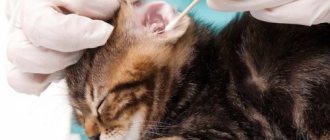
If a cat's face is swollen, the reason for this may be a number of different factors, some of which are quite life-threatening for the pet. It is not always possible to notice a tumor in time, especially when the cat is long-haired. However, once the problem has been identified, it is important to visit a veterinary clinic as soon as possible to determine the cause of the lump.
Causes and accompanying symptoms
Traumatic injuries
A cat that roams freely on the street is not protected from fights. A pet can receive various injuries, but often it is the face that suffers. You can often notice that a cat’s nose is swollen, the bridge of its nose and ears are torn, and its lips are bleeding. A broken jaw is also possible, causing the entire face to swell. When there is an open wound, it can become infected, which can lead to serious complications for the entire body.
If your pet gets into a fight, it is important to provide first aid and disinfect the wounds as soon as possible.
Neoplasms
When a pet develops a tumor on its face, it looks like a lump. Often, compression of the lymphatic vessels occurs, which causes stagnation of lymph, leading to swelling. A lump that has formed on one side of the muzzle can be not only benign, but also malignant. In such a situation, pets will require urgent surgical intervention, since cancer can cause death. In addition to swelling of the muzzle, the appearance of neoplasms provokes the following symptoms:
The appearance of neoplasms, both malignant and benign, is accompanied by diarrhea and constant vomiting.
- diarrhea;
- constant vomiting;
- bleeding of unknown nature;
- non-healing wounds.
Abscess in cats
It is the appearance of cavities in the tissues, inside of which there is pus. Often a pathological condition occurs due to infection. When the face swells, the first thing the owner needs to do is to examine the oral cavity, since cats often suffer from gumboil. It is important to remember that only a veterinarian should open an abscess. When an abscess occurs, you may notice the following symptoms:
- the area that is swollen is hot to the touch;
- when touching the tumor, the cat experiences pain and meows heavily;
- the hair on the cone falls out;
- blood and pus are released from the wound;
- general body temperature increases;
- the cat becomes apathetic.
Bites and swelling
Cats are most susceptible to bee and wasp stings. Despite the caution of pets, sometimes they can interact with these dangerous insects for fun. Often, the owner may notice that the cat's nose is swollen. The eyelids and lips also suffer. In addition to the appearance of swelling, your pet's body temperature may increase. A playful and hunting character can make a cat fight with snakes. Since the venom of these reptiles is toxic, it causes severe allergies, which manifests itself in swelling of the muzzle.
Acne in cats
In most cases, cat acne is located on the chin and, in its advanced form, causes severe swelling.
This pathology occurs not only in humans, but also in animals that also have sebaceous glands. The largest number of them are localized on the chin. Initially, the development of acne is accompanied by the appearance of blackheads. When the owner overlooked this symptom and appropriate therapy was not carried out, folliculitis occurs. This pathology is characterized by the appearance of voids with pus in the follicles. At the same time, the cat's muzzle becomes very swollen, the pet becomes less active, and its temperature rises.
Allergic reactions
Allergies in pets can be caused by chemicals, products, individual components of medications or complementary foods, pollen, hygiene products, and litter for the tray. In addition to the swelling of the muzzle, the following symptoms are also observed:
- the appearance of redness, rashes and peeling on the skin;
- itchy skin;
- sneezing;
- vomit;
- dyspnea;
- increase in body temperature.
Lymphadenitis
If your pet behaves apathetically, you should palpate the lymph nodes, as their enlargement indicates an inflammatory process.
It is an inflammation of the lymph nodes, which is not considered a separate disease, but serves as a sign that inflammatory processes are occurring inside the body. This pathological condition can be suspected if the owner closely monitors the condition of the pet. The lymph nodes that have been affected become hot to the touch. The cat becomes apathetic, spends most of its time in a lying position, loses its appetite, and at the same time absorbs copious amounts of water.
Diagnostics
In most cases, fluid accumulation occurs gradually and there is no emergency. Ascites is easy to detect, but it is not always easy to quickly diagnose what is causing it. Often, proper examination of the animal, basic blood tests and assessment of ascites fluid play an important role in diagnosis; they can provide direction for further necessary diagnostic methods.
Abdominocentesis is the removal of a sample of ascites fluid using a syringe and needle. The resulting liquid is sent to the laboratory for analysis. This may be the single most important diagnostic test for an animal with ascites because... the liquid has specific characteristics for different diseases. Ascites fluids are classified into three different categories based on cell counts and protein concentrations.
Transudates are fluids with low cell counts (less than 1500 cells/µl) and low protein concentrations (less than 2.5 g/dl). For example, transudates are formed due to hypoproteinemia, liver disease, certain tumors and blockage of lymphatic drainage.
Modified transudates are fluids with a higher content of cells (1000-7000 cells/μl) and protein (2.5-7.5 g/dl). Examples of this include transudates that form in heart failure, abdominal tumors, blockage of the hepatic vein or thoracic caudal vena cava, and some liver diseases.
Exudates are fluids with the highest cell content (greater than 7000 cells/μl) and protein concentration (usually greater than 3.0 g/dl). For example, ascites due to bleeding, tumors, feline viral peritonitis (FIP), bacterial infections that are accompanied by dysfunction of the gastrointestinal tract, chylous ascites (lymph in the abdominal cavity), leakage of urine and bile, and pancreatitis.
The pathologist also determines cell types under a microscope. Different types of cell populations indicate different pathological processes, so a cytological conclusion is necessary to make a correct diagnosis.
General blood analysis. A complete blood count (CBC) shows the ratio of red and white blood cells. Changes in the number and shape of white blood cells may indicate peritonitis. A decrease in red cells is a sign of anemia. Potential causes of anemia are acute blood loss or chronic wasting. The number of platelets (blood cells involved in clotting) is also assessed. A significant decrease in them can lead to hemorrhages (bleeding).
Biochemical research reflects the state of the body systems. In this case, it is necessary to check whether the serum albumin has decreased (hypoalbuminemia). Insufficiency of kidney function is expressed in increased levels of urea nitrogen and creatinine in the blood. Liver disease can be suspected if ALT, AST, and alkaline phosphatase are elevated. A decrease in urea nitrogen, albumin, cholesterol and, occasionally, glucose may indicate decreased liver function.
A urine test can evaluate kidney function. You should check for protein excretion in the urine (proteinuria); If the ratio of protein and creatinine in the urine is disturbed, it is recommended to check it for proteinuria and determine the amount of protein in the urine.
A chest x-ray can confirm the presence of heart or lung disease. An enlarged heart or fluid in the chest cavity may suggest right heart failure. Masses may also be found in the diaphragm, which may compress the caudal vena cava. An abdominal x-ray helps determine the size of the liver and kidneys, as well as detect any abdominal masses. Unfortunately, when ascites is large in volume, structures in the abdominal cavity are often difficult to distinguish due to the presence of fluid.
Bile acid measurement is a test specific to liver function. If ascites occurs due to liver disease, the level of bile acids increases significantly.
Measuring the level of lipase in the blood serum allows you to determine the presence of inflammation of the pancreas. This can occur with pancreatitis, cancer or abscess of the pancreas.
Abdominal ultrasound is the leading diagnostic method for ascites. First of all, fluid in the abdominal cavity enhances the image, which allows visualization of abdominal masses, changes in the liver, kidneys, spleen and pancreas. If indicated, it is possible to take a biopsy of the abnormal organ to make a final diagnosis.
If heart disease is suspected, an echocardiogram (ultrasound of the heart) is necessary. The heart valves and myocardium are visualized and cardiac function can be assessed. Heart failure can occur for many reasons, and an echocardiogram is the most informative diagnostic method that allows you to predict and prescribe treatment for this pathology.
Endoscopy is a good, relatively non-invasive method used for gastrointestinal diseases. It allows visualization and biopsy of the inner lining of the stomach and therapeutic abdominocentesis of the duodenum. Neoplasms, inflammation or lymphangiectasia (dilation of the lumen of the lymphatic vessels) of the intestine can also be diagnosed as causes of protein loss (protein release through the gastrointestinal tract).
What to do?
Help for your pet is directly related to the cause that caused the swelling of the face. If swelling occurs due to traumatic injuries, the owner should immediately apply a cold compress to the area of injury. With its help it will be possible to relieve swelling and relieve pain. Then the owner needs to take the animal to a veterinary clinic, where an x-ray will be taken to confirm or rule out a possible jaw fracture.
Tumors that occur on the bridge of the nose, in the nose, on the cheeks, chin and other parts of the face begin to be treated after diagnostic measures are carried out. First of all, a biopsy is prescribed to show the nature of the neoplasm: malignant or benign. If a growth of the first type is diagnosed, surgery is mandatory.
An abscess is treated by opening it. If the situation is advanced and the purulent cavity is greatly enlarged, surgical intervention under general anesthesia may be required. After surgery, your pet will face a long and painful recovery period. Every day, the edges of the wound will need to be moved apart to clean the cavity of secretions and put the ointment prescribed by the veterinarian into it. Therapy for allergic reactions involves identifying the allergen and avoiding contact with it. Sometimes the animal may also be prescribed corticosteroids. However, medications from this group have a number of serious side effects, so they are used only in severe situations.
Treatment
In the meantime, symptomatic treatment is necessary, especially if the case is serious and the general condition of the animal suffers. The following symptomatic therapy may be used for some (but not all) animals with ascites. It can reduce the intensity of symptoms and make life easier for your pet. However, this non-specific treatment is not a substitute for definitive treatment that addresses the disease underlying the animal's current condition.
The most important aspect of treating ascites is to determine how quickly the ascites is developing and the clinical condition of the animal. If ascites develops slowly and the animal is strong enough, then emergency care is not required. If ascites develops quickly, which is often associated with loss of strength in the animal, emergency care is necessary. Treatment until a diagnosis is made may include:
Therapeutic abdominocentesis. If a lot of fluid accumulates in the abdominal cavity, it can put pressure on the diaphragm, making it difficult to breathe. A needle is inserted through the abdominal wall and the fluid is drained to relieve pressure and make breathing easier for the animal. As soon as the animal feels better, the needle is removed. You cannot remove all the fluid, as this can lead to a shift in homeostasis in the body and shock.
Diuretics are prescribed to remove fluid from the body. They increase the excretion of fluid in the urine. Diuretics are more effective at removing fluid from tissues rather than from body cavities, so their effect on ascites is limited. The most popular drug is Lasix (Furosemide).
Oxygen therapy is often required to stabilize the animal's respiratory failure. Oxygen may be provided through a mask, nasal oxygen cannula, or oxygen chamber. Typically, once some fluid has been removed from the abdominal cavity, oxygen therapy is no longer required.
Rapidly accumulating ascites requires intravenous fluids to maintain tissue perfusion and prevent shock. If an animal has reduced total protein in the blood (due to low albumin), colloidal solutions (liquids whose osmotic pressure is equal to the pressure of the blood plasma) can be used to slow the progression of ascites.
For ascites due to bleeding into the abdominal cavity, transfusion of blood or blood components is used. If a transfusion is necessary, the animal is usually very weak and has a reduced hematocrit (a blood indicator that characterizes the degree of anemia).
If infection is suspected, intravenous antibiotics may be administered until a definitive diagnosis is made. When infected, ascites requires immediate treatment.
Prevention
Walking your cat on a leash will help you avoid unnecessary injuries or fights with other animals.
To reduce the likelihood of facial swelling in cats, veterinarians recommend that owners carefully monitor their pets. It is better not to let them go outside alone, which will protect the animals from fights, injuries and bites. Walking without an owner is most dangerous for cats that are over 3 months old. Their fear has already disappeared, but the kittens have not yet fully grown up to adequately assess the threats and fight back the offender.
It is important for cat owners to remember that industrial complementary foods sold in stores can cause allergic reactions. If a decision has been made to diversify your pet’s diet with a new food, it is recommended to introduce it gradually, observing the response of the animal’s body. An important preventive measure is a systematic examination of the cat, its ears, eyes, nose and mouth. You should attend routine medical examinations with your pet at a veterinary clinic, which will help identify the development of pathologies at the initial stages.
Symptoms of a tumor on a cat's stomach
A caring owner has probably already wondered what the symptoms and signs of a tumor are in a cat. These signs are not highly specific and can easily be confused with another disease. The signs can be confused with poisoning or toxicosis (if it is a female) here they are: weakness, loss of appetite, loss of former playfulness, activity, the animal prefers to lie down and rest.
What to do if the terrible diagnosis of a tumor in cats is confirmed
Of course, there is no need to panic in the first place. Animals perfectly sense the mood of the owner who is nearby. Remain calm, which will certainly rub off on your pet. You also need to undergo a large number of tests and prepare yourself for a long fight against the disease. Veterinary doctors will do everything possible, and will also show the owner all the possibilities of modern medicine, which is capable of real miracles. It is quite possible that our drugs and skilled doctors will help cure such a terrible disease as a tumor. We are responsible for those we have tamed, and therefore it is not recommended to abandon a pet with a tumor to the mercy of fate. Having suspected something was wrong, it is also important to warn household members that they should be as attentive as possible to the animal and, if possible, not feed it harmful sausage or sausages, even if you want to support your cat and treat him with his favorite treat.


