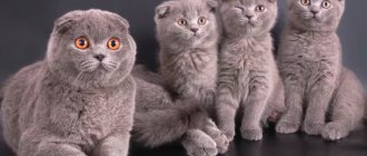
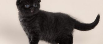
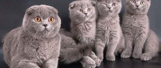
Scottish Fold cats have fans all over the world: their small, downturned ears leave no one indifferent. But, unfortunately, it is precisely because of this feature that Scottish Folds are susceptible to osteochondrodysplasia. We will find out what this disease is, how it manifests itself, and we will also look at how to care for a sick cat, what to feed and how to treat it.
Description of the disease
Osteochondrodysplasia in cats (OCD or osteochondrodystrophy) is a genetic malformation of bones and cartilage that leads to slower growth and deformation.
Interesting! Translated from Greek, “osteo” means “bone,” and “dysplasia” means “developmental disorder.”
Osteochondrodysplasia most often affects Scottish Folds, since the disease is directly related to the gene for fold ears. Scots' downturned ears remain this way due to insufficient cartilage formation and disruption of its formation. The lop ear gene is responsible not only for ear deformation, but also for disruption of cartilage formation throughout the body.
Chondrodysplasia usually affects cats' limbs, thoracic, lumbar and caudal vertebrae. This disease can also manifest itself in the form of achondroplasia. With this disease, the cat’s paws cannot grow to normal size, so the pet develops dwarfism.
Osteochondrodysplasia is not life-threatening in most cases, but it can significantly reduce the quality and duration of life.
Congenital malformations of the skeleton. Dysplasia
Due to the fact that the main molecular-biochemical defect for most OCD has not been established, etiopathogenetic treatment for these diseases does not yet exist. Therefore, the only possible option is symptomatic treatment. Since OCD are systemic diseases of connective tissue, they often also involve extraskeletal changes, which should be promptly addressed. Therefore, most patients with OCD require observation and treatment by many specialists (cardiologists, neurologists, pediatricians, ophthalmologists, etc.). An important role belongs to orthopedic treatment - both conservative and surgical.
Conservative treatment for OCD should be applied from the moment of diagnosis.
Conservative treatment can go in three directions:
- preventive treatment;
- correction of deformities;
- treatment of associated complications.
Preventative treatment
Preventive measures are effective only with early detection of OCD and consist in creating conditions for the child during growth that make it possible to slow down the pathological process.
In the first year of life, treatment should be aimed at preventing the development of deformities or their progression. In the first months of a child’s life with OCD, general muscle hypotonia, incorrect position of the head and/or torso, slight deformity of the spine and contractures of the joints of the extremities may be observed. If muscles are weak, from the first days of life it is necessary to create conditions under which the child can make more active movements. To do this, you need to put a romper on the baby and not swaddle him. Swimming strengthens muscles well. Starting from 2 months of age, the child should undergo repeated courses of general massage and constant therapeutic exercises. These procedures have a beneficial effect on muscle trophism and help increase their strength. Therapeutic gymnastics consists of passive and active movements. Passive movements maintain joint mobility. To obtain active movements in the first days of a child’s life, it is necessary to use reflex movements. So, to force the child to straighten his legs with force, you can use the crawling reflex. To do this, the child is placed on his stomach, his legs are bent at the hip and knee joints (frog pose), and his palm is placed on his feet. In this case, the child rests his feet on the hand, straightens his legs and crawls forward. All these exercises are included in the complex for practicing with a healthy child.
Subsequently, you can use gaming techniques: give the toy so that the child reaches for it with either his right or left hand; make him turn over first one way or the other; lying on your stomach, bend your torso first to the right, then to the left, etc.
In AH, in addition to general hypotension, there is looseness of the knee joints due to shortening of the femur and the convergence of the attachment points of the thigh muscles. Therefore, special attention is paid to strengthening these muscles.
A child should not be placed in pillows or taught to walk while holding hands. Such haste leads to deformation of the skeleton. After 6-7 months of life, it is better to put children on the floor and give them the opportunity to do everything they can. The child sits down and stands up only if he has sufficiently strong muscles.
For existing deformities, all therapeutic measures should be aimed at preventing their progression. If the body position is incorrect or there is a visible deformation of the spine, children should be kept in beds with a flat mattress that does not sag under the weight of the child.
With kyphosis in the thoracolumbar spine, children in the first months of life should be placed on their stomach more often and taught to sleep in this position. Lying on his stomach, the child tries to raise his head. These movements strengthen the back muscles. At an older age, the child begins to pull his legs under his body and tries to kneel, while the spine bends forward, which to some extent evens out the kyphosis. If the head position is incorrect (tilting to one side), it is recommended to place the child in a symmetrical position, placing heavy bolsters on the sides of the head (from the shoulder girdle upwards). When a child has a tilt of the head and a bend of the body that cannot be corrected by laying it with bolsters, it is necessary to make a plaster crib with the head held in a position that can be corrected. In addition, these children are recommended to undergo repeated courses of asymmetrical massage, therapeutic exercises, and swimming. During active gymnastics, it is necessary to force the child to turn in the opposite direction from his usual position. To do this, you can call him or place a toy on the side of the convexity of the curvature. After the child begins to sit down on his own, it is necessary to limit sitting as much as possible, and when this is not possible, he must be seated on a chair with a straight, high, firm back and tied to the chair so that the child does not lean forward or sideways. To do this, you need to sew a bra that secures the entire chest; Straps are sewn to the sides of the bra, which tie the child’s torso to the back of the chair.
In patients with a progressive course of the disease, preventive measures include a gentle regimen, prohibition of long walking, physical education and sports. Swimming, skiing and cycling on flat terrain are permitted.
Sanatorium treatment relieves pain, if any; the general condition improves, the child becomes more mobile. With repeated courses of sanatorium treatment, children retain mobility in the joints for a long time, contractures in the joints do not form or progress, and the progression of limb deformities in the area of the epiphyses slows down. In between courses of sanatorium treatment, exercise therapy, repeated courses of massage, and swimming are indicated.
Correction of skeletal deformities. With OCD, a large number of different deformations are observed. However, you should not try to correct every deformity.
Contractures in the elbow joints in OCD due to subluxation of the radial head usually do not respond to conservative treatment, but in the vast majority of cases they do not affect the function of the limb.
Club hand is quite easily corrected with the help of exercise therapy and the application of removable splints, which give the hand a middle position. Splints can be made from gypsum or polymer materials.
Spinal deformities, as a rule, cannot be corrected.
The greatest difficulties are presented by pathological changes in the hip joints. It must be taken into account that dislocation and subluxation of the hip in DD cannot be treated. It is usually not possible to correct a dislocation in this disease, and persistent and long-term treatment can lead to severe contractures and even greater deformation of the femoral head.
In VSED, high-standing proximal ends of the femurs are mistaken for hip dislocation due to sharp coxa vara, despite the fact that the acetabulum is well developed on the pelvic radiograph. They try to straighten the “dislocated” hip - first with splints, and sometimes simultaneously under anesthesia. This treatment results in a sharp progression of coxa vara, and the appearance of the ossification nuclei of the femoral head is further delayed. In order to avoid mistakes, you must be very careful when examining the child, and if a disproportion of the physique is detected, very carefully study the x-ray of the hip joints. If it is impossible to exclude a dislocation, then before starting treatment, contrast arthrography should be performed, which will help establish the correct diagnosis and not cause harm to the child.
Foot deformities due to OCD are treated with staged plaster casts. An exception is clubfoot in DD, as it is difficult to correct. In this case, it is necessary to start treatment from the first days of the child’s life. The treatment method is somewhat different from that for ordinary congenital clubfoot. A plaster cast is applied to the lower limb from the middle third of the thigh to the end of the fingers, but since patients have flexion contractures in the knee joints, when applying the plaster they try to straighten the limb as much as possible. When moving the foot out of a vicious position, the varus position is mainly corrected. At the same time, they try to align the equinus position, but one should not try to completely remove the foot from this position, since it is difficult to correct, and if the foot is forced out of the equinus position, a recurrence of varus deformity may occur. After the foot is moved out of the varus position and the flexion contracture in the knee joint is somewhat corrected, children are given polyvik splints up to the upper third of the thigh, with a heel. In splints, the child begins to learn to walk. Only with this disease do we recommend teaching children to walk, since they may not begin to walk at all, and our task is to give them the opportunity to move on their feet.
In some diseases (TC, Ollier's disease, etc.), asymmetric shortening of the lower extremities is observed. In these cases, it is necessary to equalize the length of the limbs. To do this, shoes on a shortened leg are “knocked”, while the front part and the heel are knocked separately in order to maintain the roll of the foot when walking. The shortening is not fully compensated, since otherwise the valgus curvature may progress. Usually, the difference in limb length is left at 0.5 cm. These children require constant monitoring, since in the first years of life the child grows quickly and the difference in limb length can change quickly.
Treatment of associated complications. Such complications include early arthrosis, acute arthritis and aseptic necrosis of the femoral head.
Early arthrosis is observed in children with severe joint deformation or when a gentle regimen is not observed. At the very beginning of the disease, rest and thermal procedures temporarily relieve pain, but due to the heavy load on the joint, they can recur. These patients are recommended for sanatorium-resort treatment, therapeutic exercises (can be done in water), and massage. Repeated courses of sanatorium-resort treatment (mud therapy, brine baths, massage, swimming, restorative treatment, etc.) delay the development of arthrosis. If left untreated, in most cases, arthrosis deformans progresses rapidly, causing an increase in contractures. The pain symptom becomes more intense and constant.
Treatment of arthritis in EDS and DD is very difficult. For ordinary arthritis, the joint is immobilized to relieve pain. Taking into account the peculiarities of the course of these arthritis, in the acute period, bed rest, slight movements in the joint (preferably in water at a temperature of 36-37°C), and painkillers are recommended. When the pain is relieved, physiotherapeutic treatment is carried out. The most effective are paraffin-ozokerite applications followed by electrophoresis of humisol.
Paraffin-ozokerite applications are applied to the affected joint (sometimes to both) at a temperature of 40-42°C. The procedures are performed daily or every other day (depending on the age and individual tolerance of the procedure to the patient). In total, up to 20 procedures are recommended for a course of treatment. Immediately after the procedure, the patient receives electrophoresis of humisol on the same joint, which is performed every other day (on the day of paraffin-ozokerite applications), also up to 20 procedures per course of treatment. Humisol can be administered from both poles. The complex of treatment measures includes injections of vitamin B12.
Aseptic necrosis of the femoral head in NSHS can be observed in MED, MPS and other diseases. Treatment is the same as for Perthes disease, but without immobilizing the joint. Unloading of the joint must be carried out until the femoral head can be restored.
Surgical treatment of diastrophic dysplasia. Surgical treatment of DD is extremely difficult, which is explained by the prevalence and severity of deformities and their steady progression.
The primary task of the surgeon is to draw up a program of multi-stage conservative and surgical treatment, the ultimate goal of which is to enable the child to move without outside support, at least with the help of crutches.
At the first stage, tissues are prepared for the intervention. The feet are corrected using staged plaster casts, eliminating varus and equinus deformities. At the same time, massage the muscles of the thighs and gluteal muscles.
The second stage is the actual surgical treatment according to the scheme developed at the CITO. It is based on intensive surgical treatment in a short time followed by long-term rehabilitation treatment.
The first operation is performed on the femoral segment. Subspinal myotomy is performed. The bone is crossed at the level of the lesser trochanter, and a wedge is selected from the proximal fragment with its base posterior and outward. The fragments are compared, eliminating flexion and adduction of the hip. If the muscle tension is strong, the femur is shortened or its proximal metaphysis is inserted like a spyglass. Osteosynthesis is performed with an extraosseous plate or an intraosseous Bogdanov nail, which is preferable, since the next osteotomy is performed in the supracondylar region of the femur to eliminate severe flexion contracture. For mild contracture, osteotomy is performed according to Repka; for severe contracture, metaplasia is performed according to Wreden. The operation is completed with plastic surgery of soft tissues according to Roux-Friedland-Volkov with the aim of open reduction of the dislocated patella or its fragmented rudiments. Thus, in one stage the vicious position in the hip and knee joints is corrected.
Correction of equinovarus foot deformity is carried out 3-4 weeks after hip surgery. The Zatsepin operation is performed, supplemented, if necessary, with an economical crescent resection of the foot. Immobilization with a plaster cast for 2 months. Then restorative treatment is carried out.
Surgical treatment of deformities on the other leg is planned similarly. The full cycle of intensive treatment lasts 5-6 months. In case of hip dislocation, only extra-articular operations are permissible.
Rehabilitation of the patient is complemented by the production of splints or lightweight orthopedic devices, orthopedic shoes. The patient is subject to long-term observation by an orthopedist.
The prognosis for the life of patients with DD is favorable.
Surgical treatment of spondyloepimetaphyseal dysplasia. For congenital spondyloepimetaphyseal dysplasia, the indications for surgery are adductor flexion contractures of the hip joints, high hip dislocations in combination with varus deformity of the neck and deformity of the hip joint.
Attempts at closed and open reduction of dislocations end in failure. Extra-articular operations are indicated: intertrochanteric osteotomies. The proximal end of the distal fragment of the femur is shifted 1-1.5 cm inward and the femur is retracted, forming an angle open outward and posteriorly at the level of the osteotomy. In the corrected position, metal osteosynthesis is performed using a bone plate. In adolescents, after osteosynthesis with the Nuzhdin-Trotsenko plate, plaster immobilization is not performed. In young children, a plaster cast is applied until fusion in the osteotomy area.
The load on the operated joint is allowed after movements have been developed. Due to the progression of arthrosis in people after 20-30 years of age, the need for endoprosthetics arises.
In patients with late spondylometaphyseal dysplasia (Kozlovsky type), indications for surgical treatment arise only when valgus deformities of the knee joint progress. Corrective osteotomies are performed according to Repka with fixation of fragments with knitting needles and plaster bandages.
For Kniest dysplasia, operations on the hip joints are performed only in adolescence, as a rule, with severe coxarthrosis with pain. More often, indications for surgery arise for clubfoot. A Zatsepin operation is performed followed by the prescription of orthopedic shoes.
Patients with metatropic dysplasia and Dyggwe-Melchior-Clausen disease are generally not operated on, the former due to respiratory failure, the latter due to mental retardation.
Surgical treatment of spondyloepiphyseal dysplasia. Surgical treatment of children with EDS, carried out in high school and adolescence, makes it possible to radically help the patient. The purpose of surgical interventions is to relieve children from pain, increase range of motion and improve the supporting function of the limbs.
Indications for surgery: flexion contractures of the hip and knee joints, dislocations of the patellas, coxarthrosis and gonarthrosis.
Flexion contractures of the hip joints are the first sign of coxarthrosis. When contracture increases, if it is not yet accompanied by a distinct pain syndrome, i.e., before the age of 10-12 years, decompression of the joint is performed by myotomy of the adductor muscles of the thigh, subspinal myotomy, and also cutting off the attachment site on the lesser trochanter of the iliopsoas muscle. In cases where the contracture is not completely eliminated, an intertrochanteric osteotomy is performed with fixation of the fragments with knitting needles, straight plates, or biopolymer clamps. The operation is performed in stages or in one stage on both sides. A plaster cast with a pelvic girdle is applied to both legs. After the cast is removed (3 weeks after soft tissue operations and 6 weeks after osteotomy), the position of the legs apart is maintained with a Vilensky splint. They prescribe classes in the pool, walking only on crutches, and exercise therapy. 4-6 months after surgery, the load on the joints is increased, but overload should be avoided.
As the child grows, contractures recur and progress, and pain in the joints appears and increases. During this period of disease development, reconstructive surgery on the proximal femur is indicated due to the progression of coxarthrosis.
The operation technique is as follows. With the patient in the lateral position, the outer surface of the femur is exposed from the top of the greater trochanter downwards using an external Langenbeck approach 12-15 cm long. Marks are made on the outer surface of the bone. At the base of the lesser trochanter, the bone is transversely osteotomized. A wedge is selected from the proximal fragment with the base outward, 2 cm high and 1-1.5 cm posterior, depending on the severity of the flexion contracture. Then osteosynthesis is performed using the Trotsenko-Nuzhdin fixator. The operation, if indicated, is complemented by myotomy of the adductor muscles and hip flexors. The leg is placed on a splint. Movements in the joint begin on the 6th-7th day. Walking on crutches is allowed after 2-3 weeks; dosed load - after 2 months and full load - 5-6 months after osteotomy. After the patient has been rehabilitated, a similar operation is performed on the other side.
Changes in the shape of the epiphyses and defective articular cartilage inevitably lead to progression of the process, and in adolescence or young age, movements in the hip joint stop. Coxarthrosis of III-IV degree develops. In these cases, total hip replacement becomes the operation of choice.
The knee joint ranks second in the frequency of clinical manifestations of EDS. These are flexion contractures that form either independently or in combination with contractures of the hip joints. In a significant percentage of cases they are caused by dislocation of the patella. In this case, the operation of choice is open reduction of the dislocation according to Roux-Friedland-Volkov.
The operation technique is as follows. A skin incision is made along the outer surface of the thigh from its upper third downwards along the outer surface of the knee joint, rounding to the tibial tuberosity. After dissecting the fascia, the rectus femoris muscle is isolated from its upper third to its attachment to the patella. The vastus lateralis muscle is cut off at the point where it transitions into a tendon sprain. The knee joint is bent and the cut portion of the quadriceps muscle is sutured to the outer surface of the rectus muscle. On the inside, the rectus muscle is sutured to the vastus medialis muscle. The attachment point of the patellar ligament is cut off together with a bone fragment and fixed with Mylar threads 1-2 cm downwards and inwards. Make sure that the patella does not dislocate when bending the tibia.
Immobilization of the limb with a plaster cast is carried out for 3 weeks. At this time, isometric tension of the thigh muscles is prescribed, and electrical stimulation of the rectus and vastus medialis muscles is performed. After removing the plaster cast, functional treatment is carried out. Physiotherapeutic procedures (paraffin applications on the knee joint), massage of the inner thigh are prescribed. Weight-bearing on the operated leg is possible after 6 weeks, and full movement is restored 6 weeks after surgery.
Congenital dislocation of the patella can be combined with valgus deviation of the tibia due to hypoplasia of the lateral femoral condyle. Under these conditions, the Roux-Friedland-Volkov operation may be ineffective and must be combined with simultaneous correction of bone deformity. From an external approach made for soft tissue surgery, the bone is freed from the periosteum in a limited area and transversely transected at the base of the condyles. By rotating the distal fragment inwards by 8-10°, a position is achieved such that the medial and lateral condyles of the femur with their anterior surface are located in. one plane. The achieved position is fixed with knitting needles, and in adolescents - with a bone plate. Movements in the joint are prescribed if there are signs of fusion.
The prognosis for EDS is favorable for life, but the patient must be under the supervision of an orthopedist all his life. Rational employment of the patient and the fight against excess body weight are especially important.
Surgical treatment of multiple epiphyseal dysplasia. Surgical interventions for MED are indicated for the development of deformity in the knee joint area, dislocation of the patellas, coxarthrosis and gonarthrosis.
In case of curvature, an osteotomy is performed in the supracondylar region of the femur or in the subcondylar region of the tibia. Osteotomy according to Repka is more often practiced. Considering the tendency to ankylosis when fixed with a plaster cast, osteosynthesis with external fixation devices is advisable. For the same reason, during surgery on the proximal end of the femur, it is necessary to use the Nuzhdin-Trotsenko plate for osteosynthesis.
Patellar dislocation in patients with MED may be accompanied by its fragmentation, which causes patellofemoral arthrosis. Therefore, it is advisable to combine the Roux-Friedland-Volkov operation with resection of the patella in the frontal plane with covering of the sawdust with local tissues.
The steady progression of arthrosis leads to the need for hip and knee replacement at a young age.
Surgical treatment of pseudoachondroplasia. Orthopedic surgeries for groin disorders are aimed at correcting deformities of the lower extremities, combating arthrosis of the hip and knee joints, and correcting short stature.
Deformity of the legs is corrected by subcondylar or supramalleolar osteotomy. Fixation of bone fragments is carried out using devices for transosseous osteosynthesis. Intervention on both legs in one stage significantly speeds up the rehabilitation process for patients.
Flexion contractures in the hip joint are eliminated by intertrochanteric osteotomy with external metal osteosynthesis; contractures of the knee joint are eliminated by supracondylar osteotomy.
Interventions aimed at correcting short stature require special care. Indications for lengthening leg segments are limited by the presence of severe deformation of the epiphyses of the bones, especially in the hip joint. Only minor extensions are permissible in case of asymmetry in leg length.
Surgical treatment of Volkoff's disease . At the initial stages, treatment is conservative: orthopedic shoes are prescribed on the healthy leg in order to compensate for the patient’s excess growth. As a preventive measure, preventive epiphysiodesis of the fingers and toes is possible. If the leg is significantly lengthened, the joint is sanitized by removing cartilaginous growths along with the patella, followed by restorative treatment. In case of cartilaginous ankylosis of the joint in combination with sharp lengthening, resection of the knee joint followed by compression arthrodesis is indicated. In the case when movements in the joint are preserved, segmental resection of the bones of the leg or femur is performed, followed by osteosynthesis with one of the external fixation devices. In case of severe damage to the knee, ankle joints, foot with severe trophic disorders, leg amputation followed by prosthetics is indicated. Growths on the bones of the skull, if they have a cosmetic defect, are removed. If cerebral symptoms increase, the patient should be treated by neurosurgeons.
The prognosis for life with damage to the skull bones is unfavorable or questionable. The disease ultimately leads to ankylosis of the affected joints.
Surgical treatment of hemimelic epiphyseal dysplasia. Surgeries should be performed in the early stages, before secondary deformities and arthrosis develop. An arthrotomy is performed, osteochondral growths are removed, and the epiphysis is carefully modeled.
Rehabilitation treatment begins 1-2 days after surgery. Physical therapy is carried out, and orthopedic shoes are prescribed if indicated.
As a rule, GED recurs. In case of relapse, a similar operation is performed in the early stages. It is preferable to carry out 4-5 economical resections without massive damage to the articular cartilage than radical operations with resection of the epiphysis bone, which always leads to severe arthrosis. With the development of segment deformity, a corrective osteotomy is performed; with severe arthrosis in adolescents, arthrodesis is performed.
Surgical treatment of dyschondroplasia. The range of surgical orthopedic treatments for dyschondroplasia is extremely wide and depends on the location and extent of bone lesions, the age of the patient, the severity of deformity and shortening of limb segments, etc.
Indications for surgery are the growth of cartilage tissue, deforming the bone and adjacent joint; deformation and shortening of a limb segment or several segments; threat of malignancy or malignancy of cartilaginous nodes.
Orthopedists are more likely to encounter dyschondroplasia in the case of damage to the metacarpal bones and phalanges of the fingers and toes. Surgical treatment for this localization is indicated for thickening of the phalanges and metacarpal bones before the development of secondary curvatures and tumor-like growths that disfigure the hand and reduce its functionality.
A staged marginal resection of the metacarpal bones and phalanges of the fingers is performed with the removal of foci of cartilaginous tissue. The resulting defects are filled with allografts. In case of relapses, the operations are repeated. This approach provides a good cosmetic result and full preservation of the function of the fingers. A similar principle is used when localizing cartilaginous nodes in the scapula and ilium, where bone resection within healthy tissue is combined with bone grafting.
Multiple deformities of the limbs with their shortening are caused by damage to the proximal and distal metaphyses, as well as the diaphysis. It is clear that it is impossible to remove all foci of embryonic cartilage due to their large number. Therefore, most orthopedic operations for dyschondroplasia are palliative in nature.
If the humerus is damaged, accompanied by its shortening and deformation, a closed correction is performed at the age of 8-10 years by applying an Ilizarov apparatus, taking into account the curvature. Closed correction is also combined with compactotomy or osteotomy through the pathological focus at the apex of the curvature. Distraction corrects deformity and compensates for shortening.
If there is a curvature in children over 10 years of age, as well as in patients with recurrent deformities, marginal bone resection is performed. The pathological tissue is then removed, the humeral axis is corrected, and the defect is filled with long cortical grafts. The Ilizarov apparatus is applied so that the proximal wires pass through the grafts, and the distal wires pass outside them. During the process of distraction in the apparatus, the distal fragment shifts along, and the formation of the regenerate sharply accelerates.
When cartilage lesions are localized in the bones of the forearm, the treatment principle is the same as when localized on the shoulder. An Ilizarov apparatus is used, in which the affected radius or ulna is lengthened by traction of the distal fragment of the bone after its osteotomy. Shortening can also be corrected using a rod apparatus.
Correction of curvatures and deformations of the femurs is carried out in the same way as for curvatures of other etiologies. Ilizarov and rod apparatuses are used.
Correction of deformities and shortening of the legs is carried out using the Volkov-Oganesyan and Ilizarov apparatuses. A combination of compression-distraction osteosynthesis with bone grafting is possible.
The peculiarity of dyschondroplasia is that relapses of shortening and deformation always develop and staged surgical treatment should be completed by the time the patient’s natural growth stops.
Dyschondroplasia is a disease in which individual nodes or several foci can become malignant at once or sequentially. Malignancy is manifested by pain and rapid growth of one of the lesions. If there is a threat of malignancy, segmental bone resection is performed within healthy tissues, followed by bone grafting. This requires close monitoring of the patient throughout his life.
Surgical treatment of metaphyseal chondrodysplasia . Surgical treatment of MCD (Schmidt, Jansen, McCuisick type, Pyle's disease, cranio- and frontometaphyseal dysplasia) is symptomatic. More often, corrective supracondylar and subcondylar osteotomies of the femur and tibia are performed. It should be taken into account that with metaphyseal dysplasias of the Schmidt, Jansen and McKusick type, deformities are prone to recurrence and require repeated corrections. The most optimal period for performing corrective operations for metaphyseal dysplasia coincides with the time of onset of ossification of the metaphyses.
Surgical treatment of mesomelic dysplasia . Indications for surgical correction of deformities are often given for dyschondrosteosis. Corrections are required for curvatures of the forearms, reminiscent of those in Madelung's deformity, as well as for varus curvatures with shortening of the legs.
Correction of deformity of the forearm and wrist joint is carried out by osteotomy of the distal metaphysis of the radius with its lengthening in the Ilizarov apparatus. The treatment technique is the same as for Madelung's deformity. Correction of deformities and shortening of the tibia is carried out by osteotomy, followed by lengthening of the segment and correction of the axis of the tibia in an external fixation device.
Correction of the shins with Nievergelt's dysplasia is more difficult, because it is combined with deformation of the epiphyses of the tibia, disruption of the shape and function of the ankle joint, and flat-valgus deformation of the feet. Such children undergo gentle osteotomies followed by transosseous osteosynthesis. Lengthening of the legs is not advisable in them, as well as in children with Robbinov's dysplasia.
Surgical treatment of mucopolysaccharidosis . Surgical treatment of lower extremity deformities is advisable only in children with preserved intelligence, i.e., with mucopolysaccharidosis type IV (Morquio syndrome).
Curvatures are corrected with subcondylar osteotomies. Operations are performed under exsanguination of the operated limb, as quickly as possible, without blood transfusions. Due to the tendency of deformities to recur in the postoperative period, a splint with a hinge is prescribed for the knee joint.
Surgical treatment of achondroplasia and hypochondroplasia . Surgical interventions for AC are aimed at correcting varus deformities of the legs and short stature.
Varus curvatures of the legs are corrected by subcondylar and supramalleolar osteotomies, using external fixation devices for osteosynthesis.
Leg lengthening with achondroplasia poses a number of problems for the orthopedist. When drawing up a long-term treatment program, it is necessary to clearly understand at what age of the child it is more rational to carry out intervention, from which segments to start lengthening, what technique to use, to know the advantages and disadvantages of all external fixation devices, to clearly know about possible local and general complications, to be able to predict the result and possible negative consequences in the long term after the end of treatment.
Lengthening the legs for a child represents a complex set of negative emotions caused by almost constant, although not intense, pain at the site of osteotomy and the insertion of wires, inconvenience in walking, self-care, and the development of a symptom complex of hospitalism. Only rare children under 10 years of age realize the need for lengthening; the majority of patients suffer from treatment. Therefore, 12 years seems to be the optimal period when leg lengthening should begin.
There are various schemes for lengthening leg segments: simultaneous symmetrical lengthening of segments, lengthening of two adjacent segments at the same time, etc. Experience dictates a differentiated approach to this problem.
In patients with a height of 135-138 cm completed at the age of 14-15 years, simultaneous lengthening of the hips is rational. In patients who are planning long-term multi-stage treatment, it is more rational to cross-lengthen according to the scheme: right thigh - left shin, then left thigh - right shin. In the interval between stages, the disproportion in the length of the segments with this scheme is little noticeable, and in the process of lengthening the patient better tolerates the presence of external fixation devices. Using this scheme, it is possible to increase height by 40-45 cm in 4-5 years, starting from the age of 10.
When lengthening the legs, it is more advisable to use the Volkov-Oganesyan, Ilizarov apparatus. Cross lengthening, especially in patients with hypertrophied muscle and fat layers, is easier to tolerate with a combination: lengthening of the tibia with the Volkov-Oganesyan or Ilizarov apparatus and lengthening of the thigh with a rod apparatus. With such a combination
Causes
Osteochondrodysplasia in cats is inherited. Its manifestation is not related to the gender of the animal.
OCD is considered a disease of Scottish Fold cats, because they are the owners of a mutant gene for fold ears. To avoid the appearance of offspring with osteochondrodysplasia, breeders always cross fold-eared cats with straight-eared cats. In this case, the risk of a dangerous gene appearing is minimal. However, in rare cases, even crossing a straight with a fold may result in a kitten with osteochondrodysplasia. That is why in some countries it is prohibited to breed Scottish Fold cats.
In Scottish Folds, the disease varies in severity. The most common disorders are the development of the bone skeleton, but in the most severe situations, deformation of the limbs can develop.
Clinical symptoms
Unfortunately, owners notice signs of osteochondrodysplasia in their pet only when they become pronounced. Main clinical symptoms of the disease:
- lameness;
- head too big;
- crooked teeth;
- stiff gait;
- jaw protruding forward;
- excessively short and flattened nose;
- the appearance of growths on the paws;
- refusal to move and jump;
- the tail is too short and inactive;
- pain during movement;
- twisted and shortened limbs;
- walking on bent legs.
- The base of the tail is too thin.
Important! Osteochondrodysplasia of Scottish Fold cats most often appears at the age of 1.5–2 months. If the gait of a small Scottish Fold is stiff and the tail is motionless, then the kitten is not recommended for purchase.
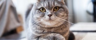
Symptoms and signs
Scottish Fold kittens are resistant to hereditary diseases, develop well, are playful and loving. However, sometimes osteochondrodysplasia affects the offspring. This hereditary disease is characterized by deformation processes in bone tissue.
Signs of the disease:
- a short tail that is not mobile;
- shortened paws with obvious signs of curvature.
Sick cats limp - this is the main symptom of the disease. Their shortened tails are inactive, sometimes becoming completely stiff. Kittens do not want to jump and run. The appearance of a “stilted” gait indicates the progression of the disease.
Most often, osteochondrodysplasia affects the breed during improper crossing, when two cats with floppy ears are brought together. A defect in endochondral ossification leads to morphological changes in the axial and peripheral skeleton, destruction of joints, formation of growths and changes in cartilage tissue.
Osteochondrodysplasia (OCSD) most often affects Scottish cats and some breeds of large, fast-growing dogs.
The disease manifests itself as difficulty in the animal's movement. It is difficult to notice this feature of a pet at the initial stage of development: with a small body weight, the cat can redistribute the load on healthy limbs. The pain syndrome provokes the pet’s refusal to run and jump. As the animal grows, it acquires an unnatural gait: diseased joints cannot withstand the load of the skeleton. Deformed limbs bend poorly, tails are shortened, and have a thin base.
The disease can be detected in the first weeks of a kitten's life. The clinical picture of the disease worsens with age.
Diagnostic methods
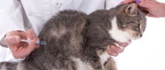
Diagnosis of osteochondrodysplasia in Scottish cats is carried out on the basis of a visual examination, as well as a study of the animal’s pedigree. In addition, to make a final diagnosis, the veterinarian will prescribe the following tests:
- General and biochemical blood test.
- General and biochemical urine analysis.
- X-ray examination of the limbs, spine and tail.
In the image of a cat with osteochondrodysplasia, you can see the deformation of the skeleton, and in the area of the limbs you can detect growths around the joints, while the gaps between them are very narrow. In severe cases of the disease, cartilage tissue is absent, and the vertebrae in the lumbar and caudal regions grow together.
Diagnostics
In order to suspect osteochondrodysplasia in a kitten, an external examination may be sufficient for a veterinarian. But to determine how far the osteoarticular deformation has gone, X-rays are taken.
The most characteristic changes affect the lower limbs, with the hind legs suffering much more than the front legs. An x-ray will show exostoses in the heel area - massive, hard osteochondral growths that resemble a tumor. The bones of the phalanges, metatarsus, and tarsal joints themselves are deformed, and the joint spaces are narrowed or absent.
Treatment
Unfortunately, there is no treatment that would permanently rid a Scottish Fold cat of osteochondrodysplasia. Typically, sick animals are given supportive care to improve their quality of life.
At the initial stage of osteochondrodysplasia, Scottish Folds are usually treated with non-steroidal anti-inflammatory drugs (Ainil, Ketoline, Meloxivet) and chondroprotectors (usually chondroitin sulfate or glucosamine). Such medications stop the destruction of cartilage tissue and increase the amount of joint fluid.
Also, as support for sick pets, manual therapy in combination with massage is prescribed. These procedures improve blood circulation in the animal’s limbs and also help preserve the structure of cartilage tissue. True, they should only be performed by a highly qualified specialist.
In the most severe cases, it may even lead to surgical intervention. Typically, Scottish Fold cats with osteochondrodysplasia undergo the following operations, which give good results:
- Osteotomy. Helps eliminate deformation and improve the performance of the musculoskeletal system, remove growths on bone tissue. For osteotomy, one has to resort to artificially breaking the bones and then connecting them in the required form.
- Arthrodesis. The joint is fixed in an optimal position, after which it gradually ossifies. Mobility in the joint is lost forever, but the support ability of the limb is preserved. Arthrodesis is usually performed if the cat is unable to move or put weight on the affected limb due to severe pain.
Interesting! In Japan and Europe, radiation therapy is used to treat cats with osteochondrodysplasia. It helps reduce pain and slow down the destruction of joints. This method is very effective, but due to the too high cost and lack of necessary equipment in Russian veterinary clinics, it is not yet available.
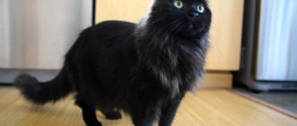
The most common diseases of Scottish Fold cats
- eye diseases; — osteochondrodysplasia; — osteochondrodystrophy; — urolithiasis Eye diseases in Scottish Fold cats
A Scottish cat can suffer from various eye diseases: inflammation of the eyelids, conjunctivitis, entropion, keratitis, etc. Let's look at these diseases in more detail. Inflammation of the eyelids. The eyelids can become inflamed due to thermal and chemical burns, injuries from the penetration of microbes, parasites and viruses. The main symptoms of this disease are: - lacrimation; - redness; - itching.
If the listed symptoms are also supplemented by thickening and deformation of the eyelid, and various types of discharge, then we can talk about inversion or eversion of the eyelid. In addition, itching, thickening and redness of the eyelids are observed with ordinary blepharitis. If the eyelid is deformed, surgical intervention is necessary; in other cases, baths should be taken and anti-inflammatory ointments should be used. The veterinarian will prescribe which medications should be used. At the first sign, you can wipe the animal’s eyes with a decoction of chamomile or calendula, as well as a decoction of green tea. Keratitis This disease of Scottish fold cats can be a transition of inflammation from adjacent tissue areas. In deep forms of the disease, the cornea may be affected. If the course of the disease is not stopped, the sclera, ciliary body, etc. are also affected. The disease must be treated immediately. Very often the Scottish Fold cat's eyes become watery. The following reasons may contribute to this: - speck. If, in addition to increased lacrimation, you also replaced the discharge of pus, then there is a possibility that a speck has gotten into your pet’s eye. In this case, you will have to resort to surgical intervention; - helminths. This is one of the most common causes of excessive tearing in cats. To eliminate this problem, purchase special anti-parasite medications; - food allergies; - infectious diseases; — lighting. If a cat stays in a room with fluorescent lamps for a long time, it may experience increased lacrimation. To avoid this, use other types of artificial lighting in the room; - characteristics of the breed. Scottish Fold cats have a flattened face, so the tear ducts may also be narrowed. This is what causes the problem of watery eyes. If the cat is still small, then there is nothing to worry about. After a few months, this problem will disappear. Otherwise, your animal's eyes need daily care. Only a doctor can determine how exactly you can help your pet. Scottish Fold cats often suffer from diseases of the musculoskeletal system: osteochondrodysplasia and osteochondrodystrophy.
How to feed sick animals
To improve the quality of life of a Scottish Fold cat with OCD, owners should make their pet’s diet as balanced as possible. It should also contain additives that would help prevent fractures and reduce the rate of cartilage destruction. A Scottish fold cat with osteochondrodysplasia can be fed both natural food and ready-made food, but veterinarians still prefer the first option.
When choosing commercial food, owners need to pay attention to ensure that it is enriched with calcium, phosphorus, iodine, iron, and vitamins E and B. You can also purchase food for your pets that was specially designed for cats with sore joints. Manufacturers additionally introduce glucosamine and chondroitin into them, which, when used in combination, enhance each other’s effects.
When eating a natural diet, it is imperative to include raw meat, sinew and cartilage in the Scots diet.
Important! There is a misconception that Scottish Folds with OCD need to be given jellied meat. However, when boiled, the required amount of nutrients does not remain in the meat and cartilage. In addition, some of them are poorly absorbed by the cat’s body after prolonged cooking.
Owners of sick cats need to remember that such animals are prone to obesity due to a sedentary lifestyle. That is why you will have to constantly monitor the calorie content of the food your pet eats, as well as your pet’s weight, and weigh it regularly. If your cat is overweight, you need to work with your veterinarian to choose a diet for her, because obesity is dangerous not only for the musculoskeletal system, but also for the cardiovascular system.
Life expectancy of cats with OCD
Of course, all owners of Scottish fold cats with osteochondrodysplasia are concerned about the life expectancy of a pet with such a disease. Fortunately, the disease does not directly affect life expectancy. When the first symptoms of OCD appear, owners should take their cat to the doctor as soon as possible. After all, timely treatment can slow down the progression of the disease and improve the pet’s quality of life.
Some veterinarians suggest that owners euthanize a sick pet or amputate the affected limb. However, with good care, supportive care and patience on the part of the owner, the condition of the Scottish Fold cat can be significantly improved. You can also increase your pet's lifespan with a balanced diet.
Unfortunately, if the disease is too severe, it is still better for the animal to undergo euthanasia. It is suggested to do it if the disease is constantly progressing, the pet does not get up at all and does not go to the litter box, and he also has to take pills that no longer bring relief. In such a situation, it is better to stop the cat’s suffering, but the decision always remains with the owner.
How can you tell if your cat is sick?
Unfortunately, animals cannot speak, so they will not be able to tell you what exactly is bothering them. The following symptoms may indicate that something is wrong with your cat:
- the animal refuses to communicate and “hides” in a corner; - rapid breathing; - the cat has lost its appetite; - if a cat experiences severe pain, then it cannot tolerate any movement and refuses to be handled; - the animal feels a constant need to scratch, lick, bite, scratch something. Very often she performs such manipulations in the place where she feels acute pain; - discharge from the eyes; — Scottish fold cat constantly runs to the toilet; - Difficulty in eating. The animal may drop food, choke on saliva, etc. If you notice any of these symptoms, contact your veterinarian for help. Before visiting the doctor, you must provide first aid to your cat (if necessary). To do this, you should have a special first aid kit for animals on hand. Never treats Scottish Fold cats with medications intended for humans.
Do straight-eared Scottish cats get sick?
Owners of the straight-eared variety of Scottish cats do not have to worry about the fact that their pets may develop osteochondrodysplasia. Scottish Straights are not susceptible to this disease, because their ears are not deformed, which means they do not have the gene for lop ears. In very rare cases, Scottish Straight cats may also experience deformation of the musculoskeletal system, but this is usually minor. This is because the Scottish cat breed is prone to various bone and joint problems.
Interesting! In addition to the Scots, representatives of the Ukrainian Levkoy breed are also susceptible to osteochondrodysplasia, since Scottish Folds were used to breed them.
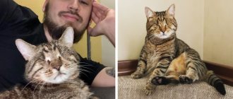
Owners of Scottish Fold cats who have been diagnosed with osteochondrodysplasia should not despair. After all, with timely treatment, proper care, supportive therapy, as well as love and care from the owner, the pet can live a long and happy life.