What are papillomas in cats and why do they appear? This disease is quite widespread among both animals and people. Why papillomas appear, how dangerous they are, and how to treat them - this is what our article is about today.
If your pussy has symptoms of illness, then do not waste valuable time, contact the Center for Emergency Veterinary Care for Animals “YA-VET”.
Veterinarians with a high level of education, training and many years of experience are at your service. Diagnostics are carried out using the latest generation equipment and with a very high degree of accuracy.
It is also possible to call a veterinarian to your home, where they will conduct an examination and make a diagnosis in a calm and familiar environment for the animal. To call for emergency veterinary help, call the phone and a doctor will be with your pet in the shortest possible time.
Reasons leading to the problem
The main source of the formation of papillomas in cats on the head, bridge of the nose, fingers, paws and other areas of the body is the papillomavirus. In every pet, like in humans, a similar pathogenic microorganism is constantly present, but in the latent phase. To activate the virus and its active reproduction, the influence of the following factors is required:
- Weak immunity. For this reason, papillomas on the nose or claw often form in kittens whose immune system is not yet fully functioning. Also at risk are sick animals, cats after giving birth and pets taking medications.
- Age-related changes in the body. Old cats often develop similar moles that need to be examined by a veterinarian.
- Heredity.
- Severe stress.
- Chronic infectious diseases.
Such growths on the animal’s body are benign. In veterinary medicine, a similar disease is also known as papillomatosis, in which small formations of a benign nature are formed. Under the influence of certain factors, papillomas in cats can degenerate into cancerous tumors, which pose a direct threat to the life of the animal. At risk of developing warts on the body are individuals living in large groups and without maintaining satisfactory sanitary and hygienic conditions. The virus is transmitted through contact of a healthy cat with a sick one, and infection is also possible through wounds on the skin. If a pregnant cat is infected with papillomavirus, then the disease is likely to be transmitted to kittens.
List and description of possible causes
It’s rare that owners bring their cat to the veterinarian complaining that it has black specks in its fur. Usually they complain of severe itching and non-healing wounds after severe scratching. If these and other symptoms are present, the following diseases should be excluded:
Demodecosis. Contrary to the erroneous opinion of some veterinarians, this disease still affects cats, although much less frequently than dogs. Demodex (Demodex cati) is considered a habitual inhabitant of the skin and normally does not cause any pathological manifestations. His activity is always provoked by something. Usually these are recent diseases of the internal organs, the postoperative period, prolonged use of antibiotics or severe hormonal fluctuations. All this manifests itself in the appearance of black grains in the cat’s fur, which are not accompanied by itching.
It is important! But demodicosis causes the appearance of large bald spots (in severe cases, even partial baldness), in place of which small black dots appear. There is also the concept of juvenile demodicosis, which occurs in kittens aged 2-6 months.
It usually affects the chin and flexor limbs. In most cases, it goes away on its own without the use of any medications. It is noted that demodicosis occurs more often in decorative breeds (Scots, British, Sphynx, Siamese). Flea dermatitis. Another reason why a cat may have black spots under its fur. A distinctive feature is that the parasites themselves may not be on the animal, since one bite is enough to provoke an allergic reaction to the flea’s saliva. The pet begins to behave restlessly, scratching its ears or chin (most often). Food allergies. A common phenomenon among decorative breeds. The slightest disturbance in the diet can provoke a severe allergic reaction, which manifests itself in the appearance of black grains in the fur, flaking, redness and hair loss (in some cases in clumps). The allergen is determined by exclusion, but most often the owners have to solve the problem for life with the help of drugs against itching and inflammation. Fleas. A real nightmare for cats of all ages. Even if you don't see them on your pet, it doesn't mean that fleas aren't biting him. You can get infected from other animals (dogs) or through objects brought from the street (even the soles of your shoes can have parasites). There are many cases when fleas were brought home along with the grass that cats love to eat in the spring and summer. At first, you may notice that your pet begins to itch more than usual (most attention is paid to the face). Over time, you may notice the appearance of black grains of sand under the fur, which fall off the animal after each stroking or combing. Large bald patches begin to appear due to scratching. In these places, brown-black crusts form on the skin due to claw wounds. The most correct solution would be to immediately treat all animals that have access to each other with anti-parasite drugs. Drops on the withers of Stronghold and Frontline are excellent against fleas and ticks. Treated 2 times a month Much attention is paid to the disinfection of housing, which is carried out using Parastop. Bacterial damage. A common occurrence in animals with a predisposition to allergies. Constant itching leads to deep openings through which infection enters. Wounds can fester and take a long time to heal. Using cytology, the pathogen is determined and effective anti-inflammatory drugs are selected. In severe cases, antibiotic therapy is used. Dermatophytosis. A fungal infection that affects the cat's fur and skin. Small black grains and redness can be found at the site of the lesion. As the process progresses, the affected area transforms into a weeping wound the size of an egg. Severe itching leads to deep scratching and the process spreading to other parts of the body. Most often, pets who have free access to the street and, accordingly, to stray animals suffer. To confirm the diagnosis, a deep pluck is taken from the lesion for microscopic examination. This area can also be illuminated under a Wood's lamp, which will show a bright green glow if there are fungal spores in the fur. Treatment is long, with the use of antifungal drugs in the form of tablets, ointments and shampoos. To avoid relapse, all places with which the infected animal comes into contact are thoroughly washed with water and bleach and chlorhexidine.
Probable locations of papillomas on the body
Characteristic symptoms of infectious papillomatosis are the occurrence of focal lesions on the mucous membranes of the mouth, the formation of pathogenic neoplasms on the outer surface of the tongue and lips. The disease is progressive and over time, papillomas can appear on the palate, pharyngeal mucosa, and nose.
At the initial phase of appearance, papules have a smooth structure. Over time, the number of lesions increases, uneven growth of the mucous membrane occurs, which provokes changes in the appearance and structure of papillomas. The number of papillomas gradually increases; in severe cases, they can cover the cheeks, gums, and can appear in the ears, on the paws, on the head, near the urethra, on the gastrointestinal tract, on the eyelids, and on the conjunctiva of infected animals.
Papillomas in the mouth cause discomfort when eating food; sick individuals may experience fever, excessive salivation, refusal to feed, bleeding in the mouth, and attacks of vomiting.
Neoplasms on the paws and ears can provoke serious inflammatory lesions, since the limbs are constantly in contact with the surface of the floor or ground, and there is a danger of ticks in the ears.
The external genitalia can also be affected; painful wounds and nodular formations are often observed here.
What species are found?
In cats, papillomas differ not only in location, but also in origin and other indicators. The table shows the main types of warts in cats and their brief description:
| Varieties | Location | Peculiarities |
| Oral | Oral cavity, tongue or inner cheek | This type of papilloma most often forms on the skin of a kitten that is less than 8 months old. |
| Oval shaped with flattened top | ||
| Multiple | Any part of the body | Diagnosed mainly in older cats and female cats |
| Many warts immediately appear, the size of which ranges from 3 mm to 3 cm | ||
| Papillomas are convex and pigmented | ||
| Single | Rarely found in felines | |
| It is a small swelling under the epidermis | ||
| Veterinarians have still not been able to figure out the reasons for the appearance of such papillomas in animals. |
Possible complications
As already mentioned, warts in animals can lead to serious consequences. Papillomatosis is often accompanied by itching. Animals scratch the affected areas and tear off tumors. Bleeding ulcers appear on the skin. This can lead to malignant cell degeneration and skin cancer.
Oncological diseases are the most dangerous, but far from the only complication of papillomatosis. When large warts are injured, severe bleeding may occur, leading to anemia. Also, sores and scratches can become infected and fester.
Symptoms
Neoplasms in cats differ in that seals appear on the body. But it is quite difficult to notice them, since usually no one probes their animal every day.
When internal organs are damaged, significant changes in the pet’s condition are noted:
- disorders of the gastrointestinal tract;
- metabolic disorders;
- neurological symptoms;
- changes in blood composition;
- respiratory problems.
Secondary characteristics:
- ascites;
- cough;
- shortness of breath, vomiting.
If a tumor in cats forms in the skin, its growth is slow. Metastasis is observed only in late stages. When a tumor grows on the mucous membranes, it grows faster and metastasizes to the lymph nodes. Melanoma in this case is prone to bleeding.
The tumor metastasizes through the blood or lymph. Those nodes that are located close to the formation are most often affected. Metastases into the dermis may be observed; they look like small dark rashes. Metastases that are transmitted through the blood can appear in various organs. The adrenal glands, brain, and liver are most often affected.
Shortness of breath may be a visible symptom of malignancy in cats over 10 years of age.
Video
More photos Author(s):
A.N.
Gerke, Ph.D., veterinary dermatologist, member of the European Society of Veterinary Dermatologists (ESVD) / A. Gerke, DVM, PhD Organization(s):
CJSC “Network of Veterinary Clinics”, St. Petersburg / “Network of veterinary” clinics", St.
Petersburg Magazine:
No. 3 - 2014 UDC 619:616.5-002:636.8
Keywords:
plasma cells, pododermatitis, cat
Key words:
plasma cell, pododermatitis, cat
annotation
Feline plasmacytic pododermatitis is a fairly rare disease of the paw pads. The clinical manifestation of plasmacytic pododermatitis is painless swelling and softening of the pads, which may ulcerate, leading to bleeding and lameness. In this case, a secondary bacterial infection develops. Laboratory tests that confirm the diagnosis include cytological and histological studies. The etiopathogenesis is not fully understood; persistent hypergammaglobulinemia in the blood serum, the presence of a plasmacytic infiltrate and a positive response to immunosuppressive therapy suggest an immune-mediated cause of the disease. There is evidence of the concomitant course of plasmacytic pododermatitis with plasmacytic stomatitis, renal amyloidosis and immune-mediated glomerulonephritis. Plasmacytic pododermatitis responds to immunosuppressive therapy, and in some cases, surgical removal of the affected tissue is performed. Spontaneous remission is also possible.
Summary
Feline plasma cell pododermatitis is a rare skin disease of cat footpads. Clinically, feline plasma cell pododermatitis begins as a soft, nonpainful spongy swelling of footpads, which in some cases become ulcerated causing haemorrhage and lameness. Recurrent haemorrhages from ulcerated or nodular areas of a pad can occur as well as secondary bacterial infection. Laboratory tests include skin cytology or skin biopsy. The aetiopathogenesis is unknown, but persistent hypergammaglobulinemia, marked plasma cell tissue infiltrate and the beneficial response to immunomodulative therapy strongly suggests an immune-mediated basis for the disease. In some cats concurrent plasmacytic stomatitis, renal amyloidosis or immune-mediated glomerulonephritis is reported. Feline plasma cell pododermatitis responds well to immunosuppressive therapy and to wide surgical excision of affected footpads. In some cases, spontaneous remission of the lesions occurs.
Etiology and pathogenesis
Plasmacytic pododermatitis is a fairly rare disease of the footpads of cats. The cause of plasmacytic pododermatitis is unknown, although the presence of elevated serum globulin concentrations, lymphocytosis, plasma cell involvement, and deposition of immune complexes in the epidermis and dermis suggests an immune-mediated nature of the disease. Gradual accumulation of plasma cells and granulation tissue leads to a soft, poorly demarcated swelling of the pad, which may later ulcerate with bleeding and secondary infection develops [7]
.
No breed, gender or age predisposition has been identified for plasmacytic pododermatitis. In one retrospective study, 61.5% of cats with plasmacytic pododermatitis were found to be infected with immunodeficiency virus (FIV), although only 19.2% had the virus detected by PCR in affected skin biopsies. [5]
.
Clinical signs
In most cats, the early manifestations of plasmacytic pododermatitis are painless enlargement and softening of the pads, which become bluish in color. The skin of the pad is dry with hyperkeratotic streaks. Primary lesions, as a rule, occur on the metatarsal and metacarpal pads, less often on the digital pads. Later, exfoliation and ulceration occur, and as the disease progresses, hemorrhagic granulation tissue protrudes and bleeds. In this case, a secondary bacterial infection may develop, and purulent-hemorrhagic, sometimes foul-smelling, exudate is released. Most often, the metatarsal and metacarpal pads are initially affected; sometimes the digital pads can also be damaged. One or more limbs may be affected [8]
.
A characteristic feature of plasmacytic pododermatitis is the absence of lameness and itching; only at a late stage of the disease, when bleeding occurs or a secondary infection develops, do cats begin to intensively lick the affected paws. Often, owners turn to a veterinarian after noticing “spontaneous” bleeding from their pet’s paws against the background of “apparent well-being.” [9]
.
Some cats may experience swelling of the nasal bridge [1]
.
In some cases, concomitant plasmacytic stomatitis was identified, manifested by bilateral proliferative and ulcerative lesions in the oral cavity. In one study, excessive salivation due to concomitant plasmacytic stomatitis was noted in 7.7% of affected cats. [5]
.
Local lymphadenopathy may be observed.
In rare cases, cats with plasmacytic pododermatitis have been diagnosed with immune-mediated glomerulonephritis and renal amyloidosis [10]
.
Differential diagnoses
Despite the characteristic clinical picture of plasmacytic pododermatitis, the list of differential diagnoses should include: trauma, contact dermatitis, burns, granulomas (eosinophilic, foreign body, bacterial, mycotic, sterile idiopathic), viral diseases (calicivirosis, rhinotracheitis, leukemia, smallpox) , neoplasms (squamous cell carcinoma, mastocytoma, lymphoma, fibrosarcoma, metastases of lung adenocarcinoma and others), autoimmune diseases.
Diagnosis is based on medical history, taking into account the nature of progression of skin lesions and response to previous therapy. The clinical examination should include examination of the entire surface of the body. During the examination, attention is paid to the presence of lesions on the skin of the fingers, interdigital space, pads, claw ridge and claws, and the oral cavity is also examined.
History data can help determine a list of differential diagnoses.
A very important clinical sign is the presence or absence of itching. The presence of itching during pododermatosis in a cat should suggest allergies (hypersensitivity to air or food allergens, insect bites), one of the clinical forms is a complex of eosinophilic granulomas, parasitosis (notoedrosis, etc.), viral and postviral dermatoses (caused by herpesvirus, calicivirus and poxvirus, postherpetic erythema multiforme), specific bacterial infections (nocardiosis, etc.), deep mycoses (histoplasmosis, sporotrichosis), as well as skin lesions associated with behavioral disorders. Calicivirus infection can cause swelling and necrosis of the footpads, but these skin lesions are usually accompanied by respiratory symptoms and fever.
In the absence of itching, dermatophytosis and tumors must be excluded. Erythema and peeling are known to often accompany allergies and dermatophytosis. Erosive lesions are also observed in allergies (eosinophilic plaques), contact dermatitis, ulcerative lesions in some autoimmune diseases, specific bacterial infections (nocardiosis, actinomycosis), deep mycoses, drug skin reactions, viral infections, epidermolysis bullosa and tumors. Crusts are often observed with notoedrosis, pemphigus foliaceus, and specific bacterial infections.
Other immune-mediated diseases, such as pemphigus foliaceus and systemic lupus erythematosus, often involve scaling and crusting, but these lesions are rarely limited to the central pad. Chemical and thermal burns most often affect the finger pads and surrounding tissues. Xanthomatosis and superficial necrolytic dermatitis can be caused by diabetes mellitus. Cases of cutaneous xanthomatosis of the footpads have been reported in cats with idiopathic hyperlipidemia. Sterile pyogranulomas and neoplasms rarely involve multiple pads, however, when one paw is affected, these differential diagnoses should be considered. Cases of metastatic digital lesions from asymptomatic pulmonary adenocarcinoma are characterized by nodular, ulcerated, painful single or multiple lesions that are often accompanied by onychomadesis. X-ray examination reveals pronounced osteolysis of the phalanges of the affected fingers and neoplasms in the lungs. Biopsy of affected fingers reveals tumor tissue, tubular structures and strands of prismatic epithelium and pseudostratified ciliated epithelium derived from the lung mucosa [4]
.
Diagnosis
Clinical examination and history play a key role in making a diagnosis, which is confirmed by cytological or histological studies.
Cytological examination of aspirates obtained with a thin needle reveals infiltration with plasma cells, including binuclear cells; mitotic figures and Russell bodies may be encountered. Neutrophils and macrophages may be present in the material obtained from ulcerative lesions [5]
. When complicated by a bacterial infection, microorganisms are detected, including phagocytosed ones.
The histological picture is characterized by superficial or deep perivascular lymphoplasmacytic dermatitis with signs of diffuse inflammation [6, 8, 10]
.
Since immunosuppressive therapy is used in the treatment of cats with plasmacytic pododermatitis, it is recommended to exclude viral and fungal infections and assess blood glucose levels before starting therapy.
Ruling out dermatophytoses should be a routine investigation performed for pododermatosis in cats. The first step is examination under Wood's lamp, despite the fact that the sensitivity of this method is low, and only 50% of Microsporum canis
. Direct microscopy of scales and fur is then performed, as well as fungal culture.
Treatment
The course of treatment is usually at least 1-2 months. To treat cats with plasmacytic pododermatitis, systemic glucocorticoids are used (oral prednisolone 1-4.4 mg/kg every 24 hours; if there is no effect, oral triamcinolone 0.4-0.6 mg/kg every 24 hours or dexamethasone 0.5 mg/ kg). When a therapeutic effect is achieved, the dose is reduced and alternative days are used. [8]
.
In case of bacterial superinfection, antibacterial therapy is justified.
The use of doxycycline at a dose of 10 mg/kg per day has shown positive results in several studies [1, 2]
. Complete remission (observation period 1 year) was observed with surgical removal of pathological tissue of the pads in combination with antibacterial therapy
[8]
.
Chlorambucil or chrysotherapy have been reported, but their effectiveness is inconsistent [10]
.
With a mild course, spontaneous remission is possible.
Literature
1. Bensignor E., Merven F. Nasal plasma cell dermatitis in cats / Veterinary Dermatology. Volume 22, Issue 3, June 2011, p. 286.
2. Bettenay SV, Mueller RS, Dow K. Feline plasmacytic pododermatitis – A prospective study of a novel treatment using systemic doxycycline. Proceedings of the 16th annual AAVD-ACVD meeting, Norfolk (USA). 4-8 April 2001.
3. Broek A., Horvath-Ungerboeck C. Pedal dermatitis in dogs and cats: Part 1 / Companion Animal. Volume 16, Issue 1, January / February 2011, p. 39-47.
4. Estrada M., Lagadic M. Feline plasmacytic pododermatitis / Pract. Med. Ch. Anim. Comp. 27(1992), p. 791-795.
5. Guaguere E., Prelaud P., Degorce-Rubiales F., Muller A., Hubert T., Lebon S. FC-23 Feline plasma cell pododermatitis: a retrospective study of 26 cases / Veterinary Dermatology. Volume 15, Issue Supplements!, August 2004, p. 27.
6. Guaguere E., Hubert B., Delabre C. Feline Pododermatoses / Veterinary Dermatology. Volume 3, Issue 1, March 1992, p. 1-12.
7. Nuttall T., Harvey RG, McKeever PJ A Color Handbook of Skin Diseases of the Dog and Cat / Second edition. Minnesota, USA.2009. P. 300.
8. Pereira PD, Faustino AMR Feline plasma cell pododermatitis: a study of 8 cases / Veterinary Dermatology. Volume 14, Issue 6, December 2003, p. 333-337.
9. Taylor JE Schmeitzel LP Plasma cell pododermatitis with chronic footpad hemorrhage in two cats / J. Am. Vet. Med. Assoc. 1990, 197, p. 375-377.
10. Vail DM Plasma cell neoplasms. In: Withrow SJ, Vail DM, eds. Withrow and Mac Ewen's Small Animal Clinical Oncology. 4 (2006). Philadelphia, PA: WB Saunders Company. p. 769-784.
Clinical case
A five-year-old British Shorthair cat presented to a dermatologist with signs of pododermatitis for over eight months. A short course of antibacterial therapy (Sinulox 50 mg twice a day for 7 days) and local treatment (levomekol, ichthyol, Yam ointments, brilliant green alcohol solution) did not bring results; on the contrary, the right metatarsal pad became ulcerated and began to bleed. Recently he began to intensively lick the ulcerated pad. Flea treatments have not been carried out for the past few years.
Clinical data and test results
The cat is vaccinated, tests for feline immunodeficiency (FIV), viral leukemia (FeLV) are negative (ViraCHECK, Synbiotics, San Diego, CA, USA), scrapings and cultures for dermatophytes are negative.
A clinical blood test revealed neutrophilic leukocytosis (band neutrophils 0.68x10⁹/l, normal 0.1 - 0.5; segmented neutrophils 15.97x10⁹/l, normal 3.2 - 9.6), monocytosis (1.21x10⁹/l, norm 0.1-0.9).
In the biochemical analysis of blood serum, pronounced dysproteinemia was noted against the background of hyperproteinemia (total protein 92.4 g/l, norm 58-80), hypoalbuminemia (18.8 g/l, norm 28-42), hypergammaglobulinemia (58.1 g/l) was detected. l, norm 13-22 g/l). The concentrations of urea, creatinine, glucose, bilirubin, ALT and AST activity are within the reference values.
On examination: the cat is of average fatness. The mucous membranes of the oral cavity are pink, clean, the lymph nodes are not enlarged. The coat is shiny. Metatarsal pads are enlarged, softened, and bluish. On the right paw there is bleeding granulation tissue measuring about 1.5 cm. The cat tries to lick this paw at the reception. The periphery of the pads is covered with crusts. Metacarpal pads are swollen, dry, with hyperkeratotic areas. The cat's front paws do not bother him.
Based on the history and clinical picture, a preliminary diagnosis of “plasmacytic pododermatitis” was made, to confirm which material was selected for cytological examination by aspiration. In cytological preparations, a large number of plasma cells were noted, which is characteristic of this disease.
Treatment and follow-up
Prednisolone was prescribed as an immunomodulator at a dose of 2 mg/kg once a day for 10 days, followed by increasing intervals between drug administration.
Enrofloxacin (Enroxil) was used as antimicrobial therapy at a dose of 10 mg/kg once a day. Dressings with Balsam of Peru were used topically. At the follow-up appointment 9 days later, the bleeding stopped and the cat stopped trying to lick the affected paw. However, general depression, polydipsia and polyuria appeared. Hyperglycemia was detected (blood glucose 13.2 mmol/l). Doxycycline (Unidox-solutab) was prescribed as an immunomodulatory drug at a dose of 5 mg/kg twice a day. Upon subsequent examination (on the thirtieth day of treatment), a significant decrease in the pathological tissue of the pad was noted; crusts were present along the periphery of the metatarsal pads. The cat is active, the appetite is normal, the glycemic level was 5.6 mmol/l. Treatment with doxycycline at a dose of 5 mg/kg twice daily was continued for another 4 weeks, and essential fatty acids in Essential-6 were prescribed topically. When examined 3 months after the start of treatment, all the pads are of normal size and consistency. A year after the end of treatment, according to the owner (by phone), there are no complaints, stable remission. Back to section
Characteristic symptoms
The appearance of such formations on the body of a fluffy can affect the quality of his appetite. Papilloma can form on the paws, stomach and any other parts of the body, including on the pad of a cat’s finger. In some pets, the formation looks like an ordinary mole; in other animals, there may be a growth that rises greatly above the skin and can cause discomfort to the cat. Alarming symptoms and pathological growths appear immediately after infection with weak immunity or after 2 months. When a cat has a wart, the following signs are additionally recorded:
- problems with appetite or complete refusal to eat;
- indifferent attitude towards everything;
- the appearance of papillomas of different locations and sizes;
- itchy sensations.
With papillomatosis, owners should monitor the cat so that he does not scratch problem areas, since open wounds will soon appear, which can become infected, and the disease will become more complicated.
Does papilloma always appear under the influence of a virus?
There is a version that the formation of warts is not always associated with the spread of viruses. This assumption sometimes turns out to be correct. Similar tumors can appear in old cats or in animals that have suffered a serious illness - in both cases, the matter is a weakening of the body’s defenses and a decline in immunity. It’s worse when these tumors are initially malignant. Sometimes the root of the problem is of a hereditary nature and lies in the presence of autoimmune diseases, which, according to experienced breeders, is confirmed in practice: papillomas present in parents quite often appear in their offspring.
Still have questions? You can ask them to our site's in-house veterinarian in the comment box below, who will respond to them as soon as possible.
Treatment of warts in cats
The most common is for your cat to eliminate the warts on its own, since cats usually develop immunity to the virus that causes warts. In this case, surgery to remove the tumor will not be required.
However, there are exceptions. Firstly, in the case of warts that do not go away on their own within about two months, surgical removal may be recommended to avoid the situation dragging on and potentially having consequences for the cat.
Secondly, in older cats or cats suffering from health problems, removal of warts may also be recommended, as the appearance of warts is usually associated with possible squamous cell carcinoma or Bowen's disease in cats.
Sources:
- https://espanol.cdc.gov/enes/flu/other/flu-in-cats/h7n2-cat-faq.html
- https://www.americanveterinarian.com/journals/amvet/2018/feb February2018/understanding-skin-disease-in-cats?p=2
- https://en.wikipedia.org/wiki/Fine-needle_aspiration
What is the danger?
If the owner notices such formations on the animal’s paw pads, then they need to be monitored and prevented from growing. Small growths on a cat's fingertips are not particularly dangerous. If the owners notice the active growth of papillomas, then they should immediately contact a veterinarian. When the formation becomes large, the blood supply to it improves and the risk of damage increases, which will lead to severe bleeding. Anemia develops against the background of large blood loss, and if a similar complication occurs in a kitten, it may die. There are other negative consequences for papillomas in cats:
- Inflammatory reaction. Violation is possible with minor or significant damage to the neoplasm.
- Cancer development. Although papillomas are benign growths, under the influence of certain factors they can degenerate into malignant formations.
Provoking factors
The following factors can trigger the activation of the virus:
- chronic illness;
- pregnancy and feeding of kittens;
- long-term use of medications;
- stress;
- avitaminosis;
- hypothermia;
- advanced age of the animal.
Small kittens often suffer from papillomatosis. Their immunity is still poorly developed.
It is important to remember that if papillomavirus has entered an animal’s body, it remains in the cells forever. Treatment can only reduce its pathogenicity and lead to the disappearance of external manifestations of papillomatosis. After therapy, the virus will be in a “dormant” state. It can be reactivated at any time as soon as the animal’s immunity decreases.
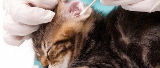
Diagnostic procedures
If a problem is detected in a cat, you need to contact a veterinary clinic as soon as possible. The specialist will examine the papillomas and find out what other clinical signs are troubling your pet. To make a diagnosis and find out the nature and causes of papillomas, the following diagnostic examinations are required:
The histological method is often used when examining an animal to determine the cause of a given disease.
- test system using immunohistochemical staining;
- histological examination;
- electron microscopy;
- PCR diagnostics.
Diagnostics
The symptoms of papillomatosis are sufficiently pronounced that diagnosis is not difficult even for a young veterinarian. Diagnosis is made based on history and examination. At the appointment, the time of appearance of papillomas, location and quantity, age of the animal, and the presence of other diseases are determined. Complex diagnostic methods are used not to make a diagnosis, but to determine the degree of development of the pathology.
Methods for effective isolation of the pathogen have not been developed, so electron microscopy, immunological and molecular methods are used to search for the virus. The role of pathological material is removed skin papillomas, as well as smears and washes of the mucous membranes.
It is easier to find the virus not in the neoplasms themselves, but in the areas adjacent to them.
The main diagnostic methods are as follows:
- Electron microscopy. They take a biopsy and make a super-thin section. After which it is stained with 2% phosphotungstic acid. The magnification is 1:100000.
- Immunohistochemical analysis. This is a reaction of hyperimmune serum with an antibody and a pathological drug with papillomavirus.
- PCR. A method for determining the genetic chains of the virus in a biopsy specimen.
How is the treatment carried out?
Medications
When cutaneous horn appears in cats, you should immediately see a veterinarian, who will conduct examinations and select therapeutic measures. In the early stages, papillomas can be treated with medication. Treatment involves the use of a number of drugs indicated in the table:
| Drug group | Name |
| Immunostimulating agents | "Kanina" |
| "Gamavit" | |
| "Maksidin" | |
| "Ronkoleikin" | |
| Ointments and solutions with antiseptic action | "Chlorhexidine" |
| "Dekasan" | |
| "Betadine" | |
| Hydrogen peroxide | |
| "Pantestine" |
Novocaine solution is often used to eliminate formations in an animal. Often, Novocain is injected into a vein to treat benign growths on a cat’s skin. A solution of 0.5-1% is suitable for therapy, with the dosage calculated at 1 ml per 1 kg of cat. The medicine is administered three times, maintaining an interval between injections of 2-3 days. It is also possible to inject Novocaine under the papilloma itself.
Surgical removal: when is it required?
Veterinarians advise that owners remove warts from cats surgically, which is more effective, and after this method of treatment there is less recurrence. Removal is performed in several ways:
- use of liquid nitrogen at low temperatures;
- laser cauterization;
- radiation.
For a speedy recovery of cats after removal of papillomas, it is recommended to give them immunostimulant vitamins and mineral supplements.
The effectiveness of folk remedies
If the virus has just begun to spread throughout the animal’s body, then the first growths can be treated with garlic juice. In the early stages, it is possible to cope with papillomatosis in cats at home, using natural ingredients. Papillomas are treated with iodine and other folk remedies are used, such as:
- fresh celandine juice;
- spurge;
- garlic juice;
- a drop of acetic acid;
- rowan berries.
Symptoms and types of papillomas
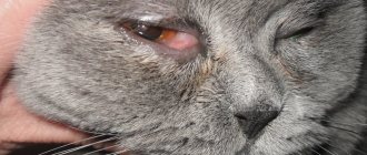
What do papillomas look like in cats? Warts are growths on the skin. They can fit tightly to the epidermis or rest on a stalk. The color of the growths can vary from flesh-colored to yellow or dark brown. The size of warts can range from 4 mm to 1 cm. In severe cases, papillomas merge and form growths similar to a head of cauliflower. A photo of papilloma in a cat can be seen below.
This viral infection is accompanied not only by the appearance of warts. Other signs of the disease are also noted:
- loss of appetite;
- lethargy, apathy;
- skin itching in the area of the rash;
- ulcers at the site of torn warts.
A general deterioration in health occurs more often in small kittens. Papillomatosis can be difficult for pups to tolerate.
Rashes can be localized in different areas of the skin and mucous membranes. Papilloma on the ear in cats most often occurs due to tick-borne infestation - otodectosis. In this case, the appearance of warts is accompanied by severe inflammation and itching in the ear. This combined disease requires persistent and long-term treatment.
Papilloma in a cat's nose can cause pain. After all, the animal’s nostrils are very sensitive. A large wart in the nasal passage can significantly complicate breathing.
Can a person become infected?
The papillomavirus, which leads to pathology in cats, does not pose a threat to the human body, since it only affects members of the cat family. Such pathogenic microorganisms are not even capable of being transmitted from cat to dog. Therefore, when the first papillomas are detected, the pet owner should not be afraid of infection, but immediately take the cat and go to the veterinary clinic in order to carry out treatment on time and prevent complications. If you are sick, you should carefully monitor the hygiene of the cat’s ears, paws and other parts of the cat’s body, and a sick animal needs good care for a speedy recovery.
Traditional methods
If fibropapilloma is a single specimen, or the owner, for example, cannot now turn to a doctor for help, he can use some traditional methods.
- The wart will disappear if the stalk of the growth is tightly tied at the very base with silk, linen or nylon thread. To make the process of eliminating the scourge faster and more effective, you can lubricate the wart with a 5% alcohol solution. The approximate time for fibropapilloma to “dry out” is a week.
- It has been proven that celandine juice can effectively cope with fibropapilloma in cats. It is necessary to find a plant and anoint the new growth with the juice of that part of the celandine that is closer to the root. There is much more juice here, it works more efficiently, and is considered the most useful. The color of the juice of this herb is bright orange. You should lubricate the wart until it decreases in size and falls off. Only the complete disappearance of the scourge can stop lubrication. Instead of celandine juice, you can use garlic juice, dandelion juice (for “young” warts), milkweed, acetic acid, and rowan berry pulp.
- It is strictly forbidden to use a chemical composition called “Clandestine” or similar to it. Look at the photo! The result of the chemical drug causes pity for the animal. Not only will he have to face incredible, hellish pain, but the scar will remain for life.
To compare the results, you can study numerous photos on the Internet and choose the best way to deal with warts.