A condition such as an eye injury in a cat is dangerous due to the loss of vision and the organ itself. There are 3 types of damage, which are accompanied by redness, deformation of the eye structures, clouding of the cornea, and swelling of the eyelids. At the first signs, the owner must take the pet to a veterinarian, who will conduct a diagnosis, prescribe medications and, if necessary, perform surgery.
According to veterinary ophthalmologist O. Fedotova, cats of brachycephalic breeds are most susceptible to injury due to short extraocular muscles and a small orbit.
Eye injury from a cat's claw
If a cat's claw gets into the eye, then several conditions are possible: non-penetrating corneal injury, penetrating corneal injury, lens injury. Each of these injuries can potentially lead to vision loss and even loss of an eye. After all, a cat's claw is a very dirty thing. Numerous bacteria live on it, there are enzymes and just particles of dirt. Because of which scratches left by a cat heal so poorly and take so long.
As soon as this entire mixture ends up in the cornea or inside the eye, the “feast” begins. Microbes actively multiply, releasing waste products that are toxic to any structure of the eye. This may lead to corneal abscess, keratomalacia, uveitis, panophthalmitis, secondary glaucoma, or ocular atrophy.
If a cat's lens is damaged, traumatic cataracts develop. And in this case, it is important to operate on such a pet as soon as possible, because the contents of the lens are foreign to the eye. It causes severe intraocular inflammation, which is already quite actively trying to “eat” the microbes left by the claw.
When all this “explosive mixture” comes to an appointment with an ophthalmologist, the matter often ends in surgery. Washing the anterior chamber of the eye, sealing the cornea, and lens surgery in cats are effective methods of minimizing the risks of complications. However, in fairness, I note that not all patients need this. An ophthalmological examination of the cat with a specialized ophthalmologist and equipment allows you to choose an effective treatment method.
Types and symptoms of eye injuries
Penetrating trauma
Occurs when sharp objects enter the organ: wood or metal sawdust, glass. And also a scratch appears as a result of a fight with relatives, incorrectly performed toilet of the eyes with poorly filtered herbal infusions. A cat can accidentally touch its eye with its claw while washing its face or, while playing, stumbles upon a sharp piece of furniture. Most often the cornea, lens, sclera, and choroids are damaged. Symptoms:
- redness and swelling;
- profuse lacrimation;
- soreness;
- unnatural squint;
- bleeding
Concussion injury
An animal can receive damage to this type of visual apparatus when falling from a height.
This type of injury is characterized by contusion of the ocular structures, in which there is no insertion of foreign bodies. Most often, a cat receives blunt trauma due to a blow with its muzzle against decorative objects, a car injury, a fall from a height, or if the owner accidentally kicks the pet. Severe hemorrhage and rupture of the sclera occurs, which contributes to the eyeball falling out of the orbit. Often, blunt trauma is combined with penetrating trauma due to crushing of the bones of the orbit and the penetration of bone fragments into the tissue of the eye. Signs:
- severe pain, the animal meows;
- swelling and hyperemia;
- displacement of the eyeball;
- tearfulness;
- swelling of the eyelids, causing the eye to be half closed;
- bleeding;
- corneal clouding;
- lack of appetite;
- depressed state.
Chemical injury
Damage to an animal's visual apparatus can be caused by ordinary household chemicals to which it has access.
Household chemicals such as alkalis and acids can injure the eye structures. The cat is damaged if the owner does not put the bottles in an inaccessible place, the cap is loosely screwed on, and a curious pet climbs in, knocking over the chemical on itself, or while the owner is cleaning the premises. Such an injury is dangerous due to melting of the ocular structures and loss of the eye as an organ. The kitten may die from painful shock. Symptoms:
- severe pain, the pet screams;
- rushing around the room in pain;
- aggression;
- the cat shakes his head;
- cardiopalmus;
- bleeding and swelling of the eyelids;
- clouding of the eye structures.
Eye contusion or blunt trauma to the eye in a cat
Eye contusion is a condition that can easily be caused by a fist fight. And a bruise to boot. As you know, cats usually fight with their paws, not their fists, but this does not prevent them from receiving concussion injuries to the eyes. In addition, injury can be caused by any blunt object: a rubber ball, the corner of a door, etc. When struck with a blunt object, the eye is deformed for some time and then returns to its original shape. Depending on the degree of deformation, either simple inflammation, or intraocular hemorrhage, or rupture of eye structures with serious consequences may develop.
Just like with a claw injury, it is very difficult to understand how serious the eye injury is in a cat. Externally, they can be almost identical. But in one case everything will go away on drops, and in the other the eye will be lost.
That is why treatment of an eye injury in a cat begins with getting the patient to the doctor as quickly as possible. I recommend that any cat with an eye injury see a veterinary ophthalmologist as quickly as possible. Despite being busy, staying at the dacha or other reasons. Find time for this. Otherwise, it makes sense to immediately accept the idea that the eye can be removed.
Eye loss
Another common injury is eye prolapse. Any animal can receive such damage, but do not forget about breed predisposition.
Traditionally, the risk group includes Pekingese, Pugs, Chihuahuas, Spaniels, Boxers and English Bulldogs. These dogs are traditionally distinguished by a short muzzle, flattened head shape and large, protruding eyes. If your pet's eye falls out, you should immediately go to the veterinary clinic. It is absolutely forbidden to correct a prolapse on your own!
It is very important that the eye does not dry out during the journey. Therefore, as an emergency measure, it may be advisable to coat the damaged visual organ with gel. And under no circumstances should there be any bandages! Contact of a bandage, like any other sterile material, with the surface of the eye in such a situation is fatal!
What is the treatment for an eye injury in a cat?
If the case is simple, then the owner will limit himself to only fear and short-term treatment with eye drops, sometimes tablets. But if, as a result of an eye injury, a rupture/displacement of the eye structures and other complications occur, the matter may end in eye microsurgery. If the degree of injury is not comparable with the future life of the eye, it is either too late - removal or prosthetic eye replacement will be the only option.
Penetrating eye injury, iris prolapse Reduction of the iris in a cat
TRAUMATIC PROPTOSIS
Displacement of the eyeball forward from the orbit most often develops in dogs of brachycephalic breeds as a result of traumatic injuries (head injury, dog bite), sometimes this pathology occurs in other dogs and cats, and may be the result of excessive restraint.
With this pathology, immediate surgical reduction is indicated to improve the preservation of the chances of vision. Preliminary abundant wetting of edematous tissues with a 40% glucose solution can reduce swelling and facilitate reposition of the eye back into the orbit. After repositioning the eye, temporary sutures on the eyelids should hold the eyelids closed for 2-3 weeks to ensure satisfactory “shrinkage” of the eye. Sutures must be placed carefully so that they do not touch the cornea and cause inversion of the eyelids. The sutures must pass exactly through the openings of the meibomian glands. Suture material – synthetic monofilament. Drug therapy should include the use of local and systemic antibacterial drugs, short-acting systemic glucocorticosteroids for a short period of time. Daily clinical monitoring to assess and stabilize associated traumatic injuries.
What to do if your cat has an eye injury?
You can help your pet by putting any antibiotic drops into the eye. It doesn’t matter whether they are human or veterinary. First, make sure that the drops do not contain hormonal substances (for example, dexamethasone, prednisolone and other anti-inflammatory drugs). If in doubt, do not drop anything until you consult with your veterinary ophthalmologist. The best treatment for your pet is to bring it to an animal eye specialist. The sooner you see a veterinary ophthalmologist, the better.
Veterinarian ophthalmologist Yastrebov Oleg.
Join us on social networks
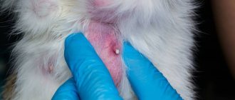
Eyelid injury
Superficial wounds of the eyelids damage only the skin and muscle layer, while penetrating wounds damage all layers of the eyelid. Since the skin of the eyelids is characterized by great extensibility and looseness of the subcutaneous tissue, swelling and hemorrhages appear here very early. The skin of the eyelids becomes tense, and the color ranges from dark blue to purple. The swelling may spread to the eyelid of the other eye.
A small external eyelid wound may conceal massive internal damage that requires immediate attention from an ophthalmologist.
Based on the appearance of the wound, one cannot draw a conclusion about the degree of damage to deeper tissues. A small external eyelid wound may conceal massive internal damage that requires immediate attention from an ophthalmologist.
If the wound is located vertically, then its edges gape due to rupture of the transverse muscle fibers. When the eyelid is injured, subcutaneous emphysema can form. This indicates a violation of the integrity of the bones of the paranasal sinuses.
Minor eyelid wounds end favorably, but if the wound becomes infected, scarring and deformation of the eyelid are possible.
If eyelid wounds become infected, scars can form, which in turn leads to cicatricial eversion of the eyelid. If the muscle that lifts the upper eyelid is damaged, ptosis of traumatic origin may appear. If you suspect the introduction of a foreign body into the tissue of the eyelids, orbit or lacrimal organs, you need to take an x-ray of the orbit.
Treatment
First aid for a wound of the eyelid - the skin around the wound should be treated with an antiseptic (miramistin, ethacridine, picloxidine, boric acid), and if the wound is contaminated, it should be cleaned and rinsed with a solution of hydrogen peroxide, and then apply an aseptic bandage.
If the eyelid wound is small and horizontal, then it does not require surgical intervention. If the wound gapes, surgical help is necessary. If it is impossible to carry out primary surgical treatment in a timely manner, it must be carried out later - even after a few days and in the absence of signs of suppuration. When treating eyelid wounds, it is necessary to take care of the damaged tissues and prevent their excision. If the wounds of the eyelids are through, then suturing is used “in two levels”: “first” - sutures on the conjunctiva and cartilage, and “second” - sutures on the skin of the eyelid.
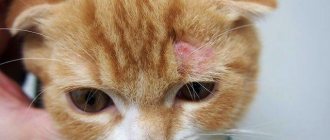
Injuries to the cornea of the eye
Most often, corneal injuries are caused by scratching with a fingernail or other foreign body, but more serious injuries, such as chemical burns, can also occur.
All corneal wounds are divided into linear and patchwork. They can be of various sizes and shapes. Clinical manifestations: lacrimation, photophobia, eye pain, blepharospasm. If an infection occurs, inflammatory infiltration of the wound edges can be detected.
To diagnose a non-perforated corneal wound, in addition to anamnestic and clinical data, the following method is used: drops of a fluorescein solution (1%) are instilled into the conjunctival sac, followed by rinsing with a sodium chloride solution (isotonic). The injured area of the cornea will turn yellow-green.
Treatment
Analgesics are not used for injury to the cornea of the eye, because this delays the healing process. Epithelization occurs within a few days without leaving a trace. If the damage was deep, it is possible that after healing there will be an area of cloudiness, which (sometimes) reduces visual acuity. In general, the prognosis for corneal injuries is favorable.
Hemorrhage into the vitreous body of the eye: symptoms and treatment
A blow to the vitreous area results in hemorrhage. Blood in the retrolental space of the vitreous expands it, and blood in the orbicular space leads to the formation of a specific rim (stripe) that surrounds the periphery of the lens behind.
Retrolental hemorrhage takes longer to resolve than orbicular hemorrhage. Sometimes minor hemorrhages may be invisible and are discovered later, when they “descend” to the lower part of the anterior chamber.
Hemophthalmos is understood as massive hemorrhage into the vitreous body, occupying a significant part of the latter.
Approximately on the third day after injury, the blood in the vitreous body undergoes a process of hemolysis with the loss of hemoglobin by red blood cells, as a result of which they become colorless and later disappear. And the hemoglobin of erythrocytes takes on the form of grains, which are subsequently absorbed by phagocytes. Hemosiderin is formed, which has a toxic effect on the retina. Sometimes the blood does not completely resolve and a blood clot is formed and replaced by connective tissue moorings. The clinical picture of hemophthalmos is dominated by loss of visual acuity from the state of light perception to complete blindness. Focal illumination and biomicroscopy make it possible to see behind the lens a dark brown granular, sometimes with a reddish tint, mass of blood that permeates the vitreous body. Ophthalmoscopy shows the absence of a fundus reflex. Later, when the blood clot dissolves, one can observe deformation of the vitreous with its liquefaction. Hemophthalmos must be distinguished from partial hemorrhage into the vitreous body, which quickly and completely resolves.
Hemophthalmos leads to the development of degenerative processes in the vitreous body.
Treatment
In case of vitreous hemorrhage, bed rest and a cold bandage are prescribed on the affected eye. Calcium preparations (tablets, eye drops and intramuscular injections), hemostatic agents (Vicasol) are used. To speed up the resorption of hemorrhage, heparin is used (subconjunctivally on days 1–2) and enzyme preparations and potassium iodide. Treatment of hemophthalmia: bed rest with the head end elevated. Use a binocular bandage for 2–3 days. Calcium chloride, pilocarpine 1% 2 times a day, glucose with ascorbic acid are used, and a solution of dicinone (12.5%) is injected subconjunctivally. Then, after 2-3 days, absorbable drugs are used: dionin, potassium iodide, lidase. Corticosteroids (under the conjunctiva) and fibrinolysin are also indicated. In the late period of treatment, ultrasound and physiotherapy help a lot. If there is no positive effect from therapy, then it is necessary to suction the vitreous, and sometimes excise part of it. The removed vitreous body is replaced with luronite (a hyaluronic acid preparation).
We must not forget that with somatic diseases (cardiovascular diseases, atherosclerosis, hypertension, blood diseases, endocrine pathology), the development of hemophthalmos is possible. But in these diseases, hemophthalmos occupies a small part of the vitreous body.
The prognosis depends on the area of hemorrhage. If the hemorrhage covers 1/8 of the vitreous body, it often resolves. With an area of 1/8-1/4 of the vitreous, moorings are formed, which leads to retinal detachment. In terms of restoring visual functions and preserving the organ in total hemophthalmia, when a blood clot occupies more than ¾ of the vitreous, the prognosis is unfavorable. Irreversible destructive changes occur in the vitreous body (organization of a blood clot, formation of adhesions). Tractional retinal detachment and atrophy of the eyeball may develop.
Types of disease
Veterinarians define three types of corneal ulcers:
- average,
- deep,
- perforative.
In the middle stage, the affected area covers the stroma. A large volume of intercellular fluid penetrates into its tissues. The cornea becomes cloudy, and partial loss of vision is observed. In the deep stage, Descemet's membrane is damaged. At this stage, the eyeball is filled with intercellular fluid. A perforated ulcer is diagnosed very rarely. If the disease is detected at this stage, the visual organ must be removed.
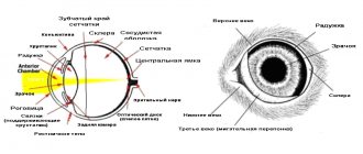
The cornea is the outer spherical membrane of the eye. Normally, it is glossy, transparent, devoid of blood vessels, and is nourished by the components of tears. It is through the cornea that we clearly see the iris and pupil. Playing an important role in the process of vision itself, the cornea serves as a barrier to microorganisms, viruses, and many drugs. As the outer structure of the eye, it is susceptible to injury and environmental influences.
The cat's cornea is glossy, transparent, spherical
The cornea of animals consists of 4 layers. From outer to inner these are epithelium, stroma, Descemet's membrane and endothelium. The transparency of the cornea is due to the fact that there are no vessels and little water in it. This result is achieved thanks to the work of endothelial cells, which are pumps that pump out fluid.
Layers of the cornea, where A – epithelium, B – stroma, C – Descemet’s membrane, D – Endothelium
Corneal injuries are classified depending on the depth of damage:
For example, damage to only the superficial epithelial layer is called erosion . It can be compared to a skin abrasion: it is superficial, very painful, heals quickly, and does not leave noticeable scars. Corneal erosions respond well to therapeutic treatment and, if they are not complicated, heal within a few days. Chronic erosions are distinguished separately. They have different causes in dogs and cats and are treated differently.
Colored corneal erosion under ultraviolet light
Damage to the epithelium and less than 50% of the stroma is called a superficial ulcer . They are also very painful, regenerate more slowly than erosions, and a slight cloudiness may remain in their place. Therapeutic treatment also has an excellent effect in the treatment of superficial corneal ulcers. Such defects can take up to 14 days to heal.
A corneal ulcer stained with fluorescein dye. An ulcer stained with a dye that glows green in ultraviolet light.
Damage to the epithelium and more than 50% of the stroma is called a deep corneal ulcer. These ulcers are less painful because there are no nerve endings in the deep layers of the stroma. However, the defect will be stained with fluorescein. Most deep corneal ulcers require surgical treatment. If the situation worsens, the deep ulcer becomes even deeper, which greatly increases the risk of perforation of the eyeball and leads to the need for surgery.
Deep corneal ulcer in a dog. Corneal edema Deep corneal ulcer in a cat. Ulcerative keratitis
Damage to the epithelium and the entire thickness of the stroma up to Descemet's membrane is called descemetocele . This is a very dangerous type of ulcer because the risk of perforation is extremely high. Descemetoceles are often not painful and do not stain with fluorescein dye, which can be confusing to the clinician. In case of descemetocele, surgical treatment of the cornea is performed, since therapy is powerless. Surgical treatment is represented by many techniques, but the most popular are transplantation of a donor cornea, one's own cornea, and a conjunctival flap. The first two techniques return the cornea to almost ideal transparency. The correct conjunctival flap will provide translucency while maintaining visual function. However, the choice of technique largely depends on the specific clinical case.
The center of the ulcer is not stained with fluorescein, since Descemet's membrane is immune to it. This confuses many clinicians.
Descemetocele may be accompanied by corneal perforation. All cases of descemetocele and corneal perforation are emergencies and require urgent surgical treatment .
The causes of erosions and ulcers of the cornea can be trauma, chemical burns, foreign bodies in the conjunctiva, entropion of the eyelids, dry eye syndrome, degenerative changes, viral, fungal and bacterial lesions. Chronic corneal erosions in dogs and cats are distinguished separately. They are caused by different reasons and are treated fundamentally differently. They will be discussed in another article.
The clinical picture of corneal damage looks like this: the animal covers its eye, there is lacrimation, photophobia, and secretion of whitish mucus. The eye itself is red (conjunctival vascular injection), the cornea at the site of injury is cloudy white or reddish white. Severe pain is present.
With deep damage, the clouding of the cornea increases (due to edema), redness of the eyeball and discharge from it persist, but pain may be less pronounced.
If you notice signs of corneal trauma in your pet, apply tetracycline eye ointment or ophthalmic gel (for example, Korneregel, solcoseryl, Vidisic) to the injured eye and take it to an ophthalmologist. The sooner you show up for an appointment, the shorter the course of treatment will be and the higher the chance of avoiding surgery.
Your pet's ophthalmologist Oleg Vladimirovich Yastrebov
Join us on social networks
Therapeutic treatment
The conservative complex includes:
- eye drops with antibacterial properties;
- antibacterial ointments;
- painkillers, sedatives;
- wearing a protective collar.
Drops of a range of antibiotics are instilled every 4 hours. You can replace them with ointments that retain the effect longer. The treatment complex includes Atropine, which relieves spasms and pain symptoms. It should be taken into account that this drug dilates the pupils and cats under its influence will experience discomfort from bright light.
Main symptoms of the disease
The symptoms of corneal ulcers are poorly identified; it is difficult even for an experienced specialist to recognize the disease at an early stage. Obvious signs appear only during periods of exacerbation; an accurate diagnosis can be established by an ophthalmological examination. The most typical symptoms include:
- increased tearfulness, moisture, shine in the eyes;
- clouding of the cornea, redness of the proteins;
- inappropriate behavior, squinting of eyes, rubbing with paws.
Symptoms of a corneal ulcer are similar to those of conjunctivitis. But this disease usually affects one eye.
General information about the disease
The cornea performs protective functions. It does not allow the external environment to influence the eyeball. The structure of this organ has three layers:
- thin epithelium;
- stroma, which is the core;
- Descemet's membrane.
At the initial stage, the epithelial layer is damaged. Access is opened for microbes and bacteria to the stroma. If the disease is detected at the stage of destruction of the epithelium, you can get rid of it in a conservative way. When the lesion spreads to the stroma, surgical intervention is usually used.