- Classification and reasons
- Symptoms of epilepsy in cats
- Diagnostic measures
- Principles of treatment
- First aid
- Drug treatment
Epileptic disease is a serious pathology of the nervous system, namely the brain. It manifests itself as seizures (or their equivalents). Epilepsy in cats is not one of the most common diseases, so its first symptoms take most owners by surprise. Awareness of the clinical picture and first aid skills during attacks is a necessary minimum for effective treatment and prevention of complications in a pet.
Classification and reasons
Epileptic seizures in cats are divided into idiopathic (primary epilepsy) and symptomatic (secondary).
Idiopathic epilepsy is congenital and is caused by genetic abnormalities. Its exact cause is difficult to determine; seizures begin at an early age and are difficult to treat.
The symptomatic (acquired) form appears in adult animals and indicates other diseases.
- Organic brain damage ranks first among the causes of secondary epilepsy in cats. This could be trauma, tumor, infectious pathology (meningitis, encephalitis), exposure to parasites.
- Chronic diseases of other organs also provoke seizures. We are talking about severe dysfunction of the liver, kidneys, and heart.
- Reaction to medications.
- Exposure to toxic substances.
- Metabolic disorders caused by improper feeding (frequent consumption of raw fish).
Secondary epilepsy in cats can be treated well (if its true cause is identified and eliminated).
Description of the disease
Epilepsy in animals is similar to a similar disease in humans.
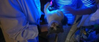
It appears suddenly: the cat, for no apparent reason, begins to worry, walk or run around the room aimlessly, then the look becomes absent, the animal falls, and begins to convulse. A clear sign is the appearance of white foam in the corners of the mouth, sometimes mixed with blood. The attack lasts no more than 5 minutes, then the animal gradually comes to its senses and returns to normal life. Epilepsy is controlled with medications. The animal will be able to lead a normal life. Regular examination by a doctor is necessary.
Symptoms of epilepsy in cats
The main manifestation of the disease is an epileptic seizure. It can proceed in different ways:
- the pet convulses (clonic convulsions) - flails its paws, jerks its head sharply;
- the cat takes an unnatural body position caused by excessive muscle tension (tonic spasms);
- the animal freezes and stares motionlessly into space.
“Classic” attacks are accompanied by the cat falling, salivation, involuntary bowel movements and urination. They last from 10 seconds to several minutes.
In most cases, seizures are preceded by a period of warning signs (“aura”). At this time, the animal becomes restless, loses orientation, makes strange loud sounds, and wants to hide. The aura lasts no more than 10 minutes.
In the post-attack period, the cat may remain disoriented, lethargic and drowsy for some time (from several minutes to several hours), or, conversely, aggressive.
If the attacks are repeated continuously and the animal does not regain consciousness, we are talking about status epilepticus. This is an emergency condition that requires qualified assistance. Epistatus threatens irreversible brain damage and even the death of the pet.
Caring for a cat during an attack
When an epileptic seizure occurs, it is necessary to protect the animal from injury during seizures, which may not respond to treatment subsequently.
- The animal's body must not be restrained. The muscles are maximally tense during an attack; restriction of movements can lead to rupture of ligaments and muscles or even fractures of the limbs.
- Turn your head to one side and place a blanket under it. This will prevent impact injuries and the possibility of saliva getting into the lungs.
- Do not put fingers or foreign objects into the mouth. The statement that an animal can develop asphyxia as a result of a recessed tongue is a myth; its muscles are tense, like the rest of the muscles of the whole body. And the introduction of foreign objects or hands into the mouth can lead to broken teeth or injury to a finger when inserted into the mouth.
- Remove any dangerous objects within the radius of the animal having an attack.
Diagnostic measures
As soon as your cat exhibits its first seizure, it should be taken to an experienced veterinarian. In addition to the examination, your pet will need a comprehensive examination.
- Laboratory tests of blood and urine.
- Electroencephalography (a specific method for studying brain potentials).
- MRI or CT scan of the brain (detects organic damage, hematomas, tumors).
- Ultrasound diagnostics of internal organs,
- ECG.
The examination is aimed, first of all, at establishing the cause of the disease. Its results make it possible to answer the question: is epilepsy in cats curable or not. If the provoking factor is not found and a diagnosis of idiopathic epilepsy is made, the prognosis for recovery is questionable.
Prevention
What should be done for preventive purposes:
- Vaccinate kittens against diseases that can cause epilepsy.
- Protect the animal from contact with poisonous and toxic substances as much as possible. Including chemicals for cleaning the apartment.
- Do not leave your pet unattended.
- It is worth protecting your pet from stressful situations. For example, collisions with a dog.
All this cannot give an absolute guarantee that epilepsy will bypass the cat, but it will help to significantly reduce the risk of developing the disease.
First aid
As soon as you notice signs of impending seizures, protect your animal by:
- remove from the window, stairs (so that the cat does not fall from a height);
- Place in a space free of objects that could cause injury.
During the attack itself, you should not interfere (the cat may scratch). There is no need to try to pull out its tongue: contrary to popular belief, the animal will not be able to choke itself with it during a seizure. It is advisable to film the course of the seizures - this is useful information for the attending physician. After an attack, provide your pet with peace and eliminate irritants (bright light, noise).
How to stop epilepsy attacks
The appearance of an epileptic attack in a cat is no reason for the owner to panic. We need to get together and provide first aid. The main actions of a cat owner during an attack:
- Laying the animal on a hard surface in a side position. It is advisable to lay something thick and soft to prevent possible head injury during an attack.
- Gently hold the animal's head with your hand, while simultaneously paying attention to changes in saliva (possible appearance of foam or blood impurities). This indicates that the animal has damaged its tongue, lip or buccal mucosa.
- At home, during exacerbation of epilepsy, doctors do not recommend self-treatment with drugs. It is not advisable to try to force a cat to drink.
It is also the owner’s mistake to use force against the animal during an attack. Trying to pin a cat to the floor during an epileptic seizure in an attempt to stop the convulsions will not achieve positive results. A cat can not only bite the owner, but also receive serious injuries. Only a veterinarian after a thorough examination can completely stop attacks or reduce their manifestation to a minimum.
Drug treatment
Treatment of epilepsy in cats with drugs must be agreed with a veterinarian. If attacks are rare (no more than one per month), medications are not required. In other cases, specific medications with anticonvulsant effects, magnesium preparations, and B vitamins are prescribed.
The dose is selected individually. The drugs are taken for a long time (in the case of idiopathic epilepsy - lifelong therapy). Unauthorized cessation of treatment is fraught with progression of the pathology.
In case of secondary epilepsy, the cause that provoked the disease is eliminated: the tumor is removed, the infectious disease is treated, and the metabolism is corrected.
The joint efforts of the owner and the veterinarian significantly improve the quality of life of a sick animal.
Diagnostics
To diagnose the disease, the veterinarian will need all available information: a description of the attacks, what medications the cat took, other diseases, treatment, etc. In addition, a test of the cat’s blood, urine, possibly an ultrasound of the abdominal organs, etc. will be required, depending on the case. .
Pediatric absence epilepsy
The effectiveness of epilepsy treatment is determined primarily by adequate diagnosis of a specific form of epilepsy or epileptic syndrome. A lot depends on the form of epilepsy or epileptic syndrome, both in the prognosis of the course of the disease (preservation of intelligence, sensitivity to anticonvulsants) and in treatment tactics. It is the nosological diagnosis that dictates how quickly anticonvulsant therapy should be started, what drugs to use, what the doses of anticonvulsants should be and the duration of treatment [1].
Childhood absence epilepsy (CAE) is a form of idiopathic generalized epilepsy, manifested mainly by typical absence seizures with onset in childhood and the presence of a specific pattern on the EEG (generalized spike-wave activity with a frequency of 3 Hz) [2]. This is one of the most studied forms of epilepsy. Despite the widespread prevalence of DAE, as well as its long-term study, the diagnosis of DAE seems simple only at first glance. There are many unresolved questions regarding both the etiology and differential diagnosis and prognosis of this disease. There are many reviews on DAE in the domestic literature. First of all, these are chapters in large monographs on epilepsy, the authors of which are M. Yu. Nikanorova and K. Yu. Mukhin [3, 4]. However, it should be noted that most reviews are based on literature data and do not take into account the introduction of video-EEG monitoring into widespread clinical practice. Currently, in the process of preparing a new International Classification of Epilepsies and Epileptic Syndromes, epileptologists are trying to formulate more clear criteria for the diagnosis of DAE. The above allowed us to once again touch upon both known and little-studied aspects of this problem.
The 1989 International Classification of Epilepsies and Epileptic Syndromes provides the following criteria for the diagnosis of childhood absence epilepsy [2, 3, 4]:
- onset of epileptic seizures at the age of 6-7 years;
- genetic predisposition;
- higher prevalence in girls;
- frequent (several to multiple during the day) absence seizures;
- bilateral, synchronized symmetrical spike waves, usually at a frequency of 3 Hz, against a background of normal underlying bioelectrical activity on the EEG at the time of the attack;
- possible development of generalized tonic-clonic seizures (GTS) in adolescence.
Below we will focus on how these diagnostic criteria are modified in the light of modern knowledge about DAE.
In the general population of patients with epilepsy, childhood absence epilepsy occurs in 2–8% of cases [2]. DAE is thought to account for 10–12.3% of all epilepsies under 16 years of age. 6.3/100 thousand - 8.0/100,000 cases of the disease are registered annually in the population of children under 15 years of age. Between 60 and 70% of all patients are girls.
Childhood absence epilepsy belongs to the category of idiopathic, i.e. those in which there is no other cause of epilepsy other than a hereditary predisposition. The type of inheritance is not precisely established. Previously, an autosomal dominant mode of inheritance with age-dependent penetrance was assumed. Later, a hypothesis about polygenic inheritance was put forward. P. Loiseau et al. consider the most likely fact that the epileptic EEG pattern and predisposition to the occurrence of epilepsy are genetically determined, and DAE itself is multifactorial and is the result of the interaction of genetic and exogenous factors [2]. It has been established that hereditary burden of epilepsy is observed only in 17–20% of patients [2]. In such cases, relatives of the proband experience both typical absence seizures and generalized types of seizures. Most cases of DAE are sporadic. AV Delgado-Escueta et al. [5] provide several loci associated with DAE (Table 1).
The table shows that only a few families with the 8q24 locus meet the classical criteria for the diagnosis of DAE, and patients with the 1p or 4p locus most likely suffer from juvenile myoclonic epilepsy with an atypical course.
However, 75% of monozygotic twins whose siblings had DAE subsequently develop the same form of epilepsy. P. Loiseau et al. believe that the risk of developing epilepsy in a child whose parents suffer from DAE is only 6.8% [2].
Clinical manifestations
It is believed that epileptic seizures in DAE can begin between the ages of 4 and 10 years. The maximum number of cases of the disease occurs between 5 and 7 years of age. The onset of this form of epilepsy before the fourth year of life is unlikely, and after 10 years it is extremely rare [2].
Type of epileptic seizure. According to the International Classification of Epilepsy and Epileptic Syndromes of 1989, two types of generalized seizures are considered characteristic of DAE - typical absences and GTCS. Recent studies have shown that these two types of seizures cannot occur simultaneously in DAE. The onset of DAE is characterized by only typical absence seizures, and GTCS can be observed in adolescence (after 12 years), when absence seizures have already disappeared.
As you know, a typical absence seizure is a short (within a few seconds) generalized type of attack.
with a sudden start and end against a background of impaired consciousness. Typical absence seizures have their own clinical and EEG features in various absence epilepsies (see differential diagnosis). The child stops purposeful activity (talking, walking, eating), becomes motionless, and his gaze becomes absent. Mild automatisms are often observed, especially in the first seconds of an attack, as well as a clonic or tonic component of an attack. Possible pallor. Urination is extremely rare. The child never falls during the attack and resumes purposeful activity a few seconds after the end of the attack.
Absence is typical for DAE and has the following characteristics:
- factors provoking an attack - hyperventilation (in 100% of all cases), emotional stress (anger, fear, surprise, admiration, grief), intellectual factors (lack of interest, absent-mindedness);
- attacks may disappear or their frequency may decrease with physical and intellectual stress;
- the duration of the attack ranges from 4 to 20 s;
- high frequency of attacks (pycnoleptic course); it is difficult to establish the true frequency of attacks without video-EEG monitoring, but several tens to hundreds of attacks per day can be observed;
- attacks, as a rule, become more frequent after waking up or in the evening;
- consciousness is completely lost (after an attack - amnesia);
- automatisms (the complex nature of absence seizures) are often observed.
According to P. Loiseu et al. (2002), clinical criteria for excluding the diagnosis of DAE are [2]:
- simultaneous presence of absence seizures and GTCS in the clinical picture;
- incomplete impairment of consciousness or preservation of consciousness at the time of the attack;
- severe myoclonus of the eyelids, single or irregular myoclonus of the head, trunk or limbs during absence seizure.
The last statement is controversial, and not all authors agree with it. At the same time, all researchers agree that the presence of a myoclonic component of a typical absence seizure, as a rule, is significantly more often observed in DAE resistant to anticonvulsants.
Data on the frequency of generalized tonic-clonic seizures in adolescents with DAE are contradictory. It is believed that they are observed in 30–60% of all cases. Perhaps such a high percentage of convulsive seizures is due to the fact that researchers do not always strictly adhere to the criteria for the diagnosis of DAE, and some of the observed patients suffered from other absence epilepsies. GTCS begins 5–10 years after the onset of absence seizures. Researchers note the fact that attacks are rare and can be easily controlled with anticonvulsants [2]. Some patients may experience isolated attacks after lack of sleep or against the background of a stressful situation. Factors that increase the risk of developing GTCS in adolescence are the male gender of the patient, relatively late onset of epilepsy (at the age of 9–10 years) and late initiation of treatment (in 68% of cases) [2].
The neuropsychic status of most patients with childhood absence epilepsy remains normal. There are no intellectual impairments [3, 4]. However, hyperactivity and/or attention deficit disorder is observed in 30–50% of children, which is significantly higher than the population frequency of 5–7%. Most patients have learning difficulties [6], which, like behavioral characteristics, can be caused by various reasons: the attacks themselves, the influence of anticonvulsants, and psychological changes observed in the patients’ parents [2].
Absence status. In some children with DAE (in 7–24% of all cases), episodes of absence seizure status may occur. At the same time, the frequency of absence seizures increases, which leads to the development of a special condition, characterized primarily by varying degrees of impairment of consciousness: from mild drowsiness to stupor and lethargy. This condition lasts from one hour to several days and often ends in a generalized tonic-clonic seizure.
Electroencephalographic characteristics of DAE
Seizure EEG. Characterized by a bilateral, synchronous, symmetrical discharge of the “spike-slow wave” complexes. The frequency of paroxysmal EEG abnormalities increases significantly with hyperventilation and during sleep (during the transition from REM to NonREM phase). Both hyperventilation and waking EEG recordings after sleep deprivation can be used to provoke an epileptic pattern. It must be emphasized that various EEG variants of a typical absence in DAE occur much more often than a certain “ideal” averaged EEG pattern described in most epileptology manuals [7].
DAE is characterized by the following features of the spike-wave complex:
- in the structure of the “spike-slow wave” complex there can be from 1 to 2 spikes;
- During the first 2 s of the attack, only polyspikes (without slow waves) are possible. This onset is usually associated with the myoclonic component of absence seizure;
- the discharge frequency ranges from 2.7 to 4 Hz;
- there is a decrease in the discharge frequency towards the end of the attack (by 0.5–1 Hz);
- the amplitude of the complex is most pronounced in the fronto-central EEG leads. The amplitude of the spike may decrease towards the end of the attack, sometimes the spike completely disappears;
- Variable asymmetry of the discharge amplitude is possible, especially in patients undergoing treatment;
- the duration of the discharge is not less than 4, but not more than 20 s (on average 10–12 s). A complex lasting more than 5 s usually has clinical manifestations;
- The epileptic discharge begins abruptly and ends gradually.
P. Loiseau et al. (2002) believe that DAE is not characterized by fragmentation (rupture) of the discharge on the EEG, the presence of multiple spikes and photosensitivity. The last statement does not seem very correct. Photosensitivity (or photosensitivity) is the appearance of polyspike-slow wave complexes on the EEG in response to photostimulation. Photosensitivity appears to be inherited separately from DAE and can be observed with it, but with a frequency not exceeding that in the general pediatric population. It is believed that the peak frequency of the photoparoxysmal response occurs between the ages of 7 and 14 years - it is observed in 14% of boys and 6% of girls. After puberty, the incidence of photosensitivity in a population of healthy people is 1%. In any case, there appears to be no benefit in subjecting children with DAE to repeated photosensitivity tests if the first test is negative. In order to prohibit a child with DAE from watching TV or playing on the computer, it is necessary to prove not photosensitivity itself, but the fact that he has photogenic epilepsy (with the development of epileptic seizures in response to a specific provoking photogenic factor). A child in such a situation should undergo thorough testing to determine the nature of the photogenic provoking factor, and it can be very different (flickering sunlight, flashing artificial light at a disco, flickering TV or computer screen, etc.).
It is assumed that the generalized spike-slow wave discharge is genetically determined, but is inherited separately from DAE [7]. Therefore, it can occur in a variety of generalized forms of epilepsy and in healthy people. The hypothesis about the autosomal dominant mode of inheritance of this EEG pattern is currently considered untenable. It is necessary to take into account the data that a generalized spike-slow wave discharge can occur in approximately 1.5-2% of children without epilepsy. As a rule, these are healthy siblings of children with epilepsy. In these cases (in the absence of clinical seizures), discharges have no clinical significance, but reflect a genetic predisposition to the development of seizures. Treatment, according to H. Doose [7], is necessary only in cases where the frequency and severity of these EEG manifestations increases over time. Therefore, the diagnosis of DAE should be made not based on encephalographic data, but solely on the basis of the totality of the clinical and encephalographic picture.
Interictal EEG in DAE is usually described as “normal.” However, some variants of disturbances in basic bioelectrical activity are possible. Quite often, parieto-occipital delta rhythms are observed, which decrease when the eyes are opened [7]. This phenomenon is regarded as a favorable prognostic symptom—such patients are less likely to experience GTCS in the future. If there is a diffuse slowdown in the main bioelectrical activity, then first of all we must exclude an overdose of drugs and the presence of valproate encephalopathy. A slowdown may also occur with inadequate treatment of DAE with phenytoin.
The EEG during absence seizures shows continuous generalized epileptic activity, often, but not always, similar to that characteristic of a single typical absence seizure.
Differential diagnosis
Differential diagnosis must be made with other forms of epilepsy, which are characterized by typical absence seizures. In addition to DAE, the “continuum” of absence epilepsies includes the following forms of epilepsies:
- juvenile absence epilepsy;
- juvenile myoclonic epilepsy;
- epilepsy with myoclonic absence seizures;
- perioral myoclonus with absence seizures;
- myoclonus of the eyelids with absence seizures;
- absence epilepsy of early childhood.
All of the above forms of epilepsy are characterized by typical absence seizures, but their clinical features, as well as the characteristics of the course, vary depending on the form of epilepsy. Absence in these forms of epilepsy can be either the only type of seizure or combined with other types of generalized seizures (GTX, myoclonus), and can be either short (6–10 s) or long (about 30 s). It can occur with either complete or partial impairment of consciousness [8, 9]. The degree of severity of automatisms and motor components of absence also varies in individual forms of epilepsy (Table 2).
A rather difficult diagnostic situation arises in cases where typical absence seizures begin before the age of 4 years. Typically, if absence seizures occur in early childhood, they are different from those with DAE. Absence seizures in this case have a spanioleptic course, can be combined with other types of seizures (myoclonus, GTCS), and are resistant to anticonvulsants. Children with this type of absence seizure usually have neurological disorders and suffer from mental retardation. Absence epilepsy of early childhood, described by H. Doose et al. in 1965 and included in the International Classification of Epilepsies and Epileptic Syndromes in 1989, is currently not considered nosologically independent. Typical absence seizures have also been described in the structure of symptomatic epilepsy with a variety of brain injuries - arteriovenous malformations, tumors, meningitis, etc. [2]. In this situation, it is still unclear whether typical absence seizures are a consequence of brain damage or an accidental combination of brain damage and idiopathic form of epilepsy.
Certain diagnostic difficulties may arise during the differential diagnosis of a typical absence seizure and a complex focal seizure. As a rule, a frontal focal seizure may clinically look like an absence seizure, since it can cause rapid spread of the attack to both hemispheres. However, the EEG shows focal changes that precede the appearance of spike-wave complexes.
Evolution and forecast of current
The average duration of absence seizures with DAE is 6.6 years, i.e. e. absence seizures disappear at the age of 10–14 years [2]. The disappearance of absence seizures does not always mean recovery from epilepsy, as GTCS may occur during puberty (see above). In 6% of all cases of DAE, absence seizures persist in adults. They become rare, sometimes occur against the background of provoking factors (lack of sleep, menstruation), and their clinical manifestations are less pronounced [2].
Patients with DAE require anticonvulsants, since epileptic seizures are very common and can cause cognitive impairment. In addition, the possibility of developing a non-convulsive status must be taken into account. The goal of treatment for DAE is to achieve clinical-encephalographic remission, i.e. a stable (at least two years) absence of clinical attacks and normalization of the EEG. According to various authors, clinical and laboratory remission against the background of anticonvulsants is possible in 75–90% of all cases [2, 3, 4].
Epileptic seizures in DAE are well suppressed by both broad-spectrum anticonvulsants (sodium valproate, lamotrigine) and anti-absence drugs (ethosuximide). On the other hand, epileptic seizures in DAE are prone to aggravation (increased frequency of attacks and development of status epilepticus) when carbamazepine or vigabatrin are prescribed. Some drugs (phenytoin and phenobarbital) are not effective in treating absence seizures [10].
It is believed that sodium valproate (Depakine, Convulex, Convulsofin) and ethosuximide (Suxilep) are equally effective in the treatment of DAE, and they are the first choice drugs [1, 2, 3, 4, 10, 11]. For DAE without generalized convulsive seizures, the basic drug for monotherapy is ethosuximide (suxilep), which is prescribed according to the following scheme: for children under 6 years of age, start with 10–15 mg/kg (no more than 250 mg/day) with a gradual increase in dose every 4–7 days until remission of attacks is achieved or the first signs of intolerance to the drug appear. In children over 6 years of age, you can start with 250 mg/kg, the maintenance dose is 15–30 mg/kg/day.
The possibility of achieving remission with monotherapy with sodium valproate and ethosuximide is 70-75%, with lamotrigine (Lamictal) - 50-60% [2]. In addition to its slightly lower effectiveness, lamotrigine has another drawback - a slow rate of administration. When using sodium valproate, preference should be given to long-acting dosage forms [11], namely Depakine Chrono. Double use of Depakine Chrono during the day ensures a stable concentration of the drug in the blood, and therefore its effectiveness and reduces the risk of side effects. In addition, a convenient dosage (2 times a day) makes it easier for the patient to comply with the drug regimen.
A small proportion of patients (10 to 15% of all cases of DAE) achieve remission when prescribed low doses of Depakine Chrono (10–15 mg/kg body weight per day). If remission is not achieved, then the dose should be increased to 20–30 mg/kg body weight per day. If necessary, increase to 40 mg/kg body weight per day, provided the drug is well tolerated. Monotherapy cannot be considered ineffective until the maximum tolerated dose of anticonvulsants has been reached. If the maximum well-tolerated doses of depakine chrono are not effective, alternative monotherapy with ethosuximide (up to 20 mg/kg body weight per day) or lamotrigine (up to 10 mg/kg body weight per day) is possible [2, 11].
If sequential monotherapies are ineffective, the use of 2 anticonvulsants is indicated [1, 2, 3, 4]. The combination of sodium valproate and ethosuximide has been proven to be more effective than monotherapy with these drugs. It is also suggested that sodium valproate and lamotrigine have a synergistic effect [1]. Therefore, this combination is possible in the treatment of DAE. The dose of lamotrigine, if combined with sodium valproate, should be reduced to 5 mg/kg body weight per day.
Resistance to anticonvulsants is observed in 5–10% of all cases of DAE. Even in the presence of such resistance, a significant reduction in the number of attacks under the influence of anticonvulsants is usually possible.
Cancellation of anticonvulsants is possible 1.5-2 years after the cessation of epileptic seizures. The optimal duration of anticonvulsant treatment for DAE is still not known; it can only be determined by conducting long-term prospective multicenter studies on this issue. The condition for discontinuation of anticonvulsants is also the normalization of the EEG. It is believed that the abolition of anticonvulsants should be carried out gradually (over 4-8 weeks) in order to prevent relapses. If repeated GTCS occurs during puberty, this serves as a reason for re-prescribing anticonvulsants.
Epileptology is a branch of neurology that is characterized by extremely rapid development. Our ideas about the etiology and course of various epilepsies and epileptic syndromes are constantly changing, and the criteria for their diagnosis are also being revised. We hope that the new classification of epilepsies and epileptic syndromes, which is currently being created, will take into account all the latest data regarding DAE.
For questions regarding literature, please contact the editor.
E. D. Belousova, Candidate of Medical Sciences A. Yu. Ermakov, Candidate of Medical Sciences, Moscow Research Institute of Pediatrics and Pediatric Surgery, Ministry of Health of the Russian Federation, Moscow
Cats at risk
Although epilepsy can occur in any cat, it most often affects:
- animals with a similar disease in their pedigree;
- kittens whose mothers suffered a serious illness during pregnancy;
- kittens obtained from close relatives;
- survivors of chemical poisoning;
- have had infections, often with complications;
- cats that have been injured or stressed during pregnancy.
There is no age interval for the onset of the disease - seizures occur in kittens and adult cats.