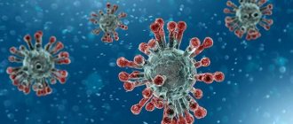
Feline infectious peritonitis (FIP) is an extremely dangerous viral disease that can be fatal. Therefore, peritonitis, along with FIV, panleukopenia and leukemia, is considered the most terrible infection. How to recognize peritonitis in a kitten and how to prevent this disease from appearing in your pet - an article from the Murkosha shelter team will help you figure this out.
1) The causative agent and features of the course of the disease
2) Risk factors
3) Routes of infection
4) Symptoms of peritonitis
5) Diagnosis and treatment
6) Prevention
Definition of disease
Feline viral peritonitis is a highly contagious disease characterized by damage to the immune system. The peculiarity of FIP is a very long incubation period and the absence of specific symptom complexes that complicate early diagnosis.
Pathogen
FIP is caused by RNA viruses, which are classified as coronaviruses. This family is large, but only two strains affect cats:
- Feline enteric coronavirus (FECV, feline viral enteritis). Manifests itself as symptoms of gastrointestinal disorders (diarrhea, vomiting). After about 3-5 days, the symptoms subside, and it seems that there is no longer an infection in the animal’s body. In fact, the cat remains a carrier of the virus for a long time and releases it into the external environment.
- Feline infectious peritonitis virus (FIPV, feline viral peritonitis). In the early stages it has no specific symptoms and begins spontaneously and suddenly. It is characterized by damage to the immune system (T-lymphocytes - macrophage cells), depriving the body of its main defense against external pathogens.
There is an opinion that the causative agent of viral enteritis (FICV) causes dry peritonitis in cats, and the causative agent FIPV causes its wet form.
Preventive measures
Feline peritonitis spreads through contact and excrement, so the main method of prevention is vaccination and cleanliness of the premises. The Primucel vaccine does not provide a 100% guarantee, but it significantly reduces the risk of infection. Such animals have a chance to cope with the infection and recover.
In order for domestic cats to live long, it is important to create appropriate conditions for them. It is necessary to provide:
- a good balanced diet with sufficient vitamins and minerals,
- calm environment,
- love of the owners,
- a clean, warm place to sleep.
It is important to monitor the hygienic condition of cat litter boxes and cat fur, and carry out timely sanitary treatments against fleas and ticks. At the slightest suspicion of ill health, you should contact a veterinary clinic.
It is best to isolate a pregnant or lactating cat if there are other cats in the house.
Early diagnosis will help identify the disease, and prompt use of medications will save your pet’s life. Thanks to preventive measures, the cat will live many happy years next to its owners.
Mechanism of disease development
The coronavirus penetrates through the villi of epithelial cells lining the oral or nasal cavity and nasopharynx. Further, pathogenesis can develop in two ways: either a dry form with initial symptoms of digestive disorders, or a wet form, which has no specific symptoms at the first stage.
In the dry form of feline infectious peritonitis, the virus can remain in the body for a very long time and not manifest itself in any way. But under certain circumstances, its virulence increases (a kind of natural passage of the strain) and its pathogenicity increases sharply.
Important! The virus can “sleep” in a cat’s body for a very long time. Perhaps this is influenced by the general condition of the body, its ability to resist pathogenic agents.
With the wet form of feline infectious peritonitis, the process develops much more dynamically, but specific visual signs appear only in the last stages.
After the virus becomes as pathogenic as possible, the immune system responds by releasing macrophages into the blood - T lymphocytes, and then B lymphocytes. These are tools of humoral immunity that are responsible for the formation of a specific immune response and nonspecific protection.
Most often, these measures are enough to overcome the disease, but there are a number of pathologies in which viral particles infect cells of the immune system. These are immunodeficiency diseases, which also include peritonitis.
In this case, the main target of the virus will always be the immune system and its “tools”. This is the only protector of a living organism that nature has endowed it with.
During a viral attack, the released macrophages do not fully perform their function - they absorb aggressive particles, but cannot kill them. As a result, the virus feeds on the reserves of the affected macrophage, destroys it and comes out unharmed.
But the pathogenic effect of the virus does not end there; it penetrates all organs and tissues. A large number of aggressive particles accumulate in places where there are many small capillaries - in the liver, spleen and other organs.
When the immune system is weakened, under the influence of the virus, the porosity of the capillaries (the permeability of the vascular walls) increases and part of the liquid fraction of the blood goes beyond their limits. Fluid accumulates in the abdominal cavity, causing ascites (dropsy) and inflammation of the peritoneum. This is how a transient wet form of peritonitis develops.
If the immune system is strong enough, it actively resists and manages to produce antibodies to the virus. Then the process of struggle is delayed, but all internal organs are also involved in it. This is the dry form of FIP.
Pathological changes
A standard complication is the accumulation of viscous clear fluid in the abdomen or chest area, sometimes with visible flakes and fibrin thread.
This same fibrin forms a film covering the tissues and membranes of internal organs. At the same time, they become dim, and mini-spikes are observed in different areas.
Moreover, whitish foci of rotting are often found there, surrounded by compacted exudate (taking the form of small nodes or plaques with a diameter of up to 10 mm). This affects the liver, pancreas, intestinal wall and other membranes through which necrosis penetrates.
In the lungs there are fewer such formations, and the pathways themselves acquire a rich scarlet color, often becoming denser.
The clinical picture also includes enlargement of the kidneys against the background of the appearance of single white nodes, absorbed into the cortical composition.
Did you know? When frightened, a cat can jump to a height 5 times its height.
With proliferative dynamics, foci of inflammation arise, covering the eyes and nerve endings, cardiovascular lines and the lower abdominal region.
Routes of infection and spread of peritonitis
A cat becomes infected through contact with a source of infection. Coronaviruses do not have significant resistance to environmental factors. After identifying an infection, it is recommended to wait 2-4 months (depending on the disinfection treatments carried out in places of detention) before getting a new animal.
The virus can be transmitted through contact and nutrition. In addition to sick animals, household items can serve as a source of infection - feeding dishes, tray, bed, and so on. Also, a sick cat can transmit the infection to kittens during birth or feeding.
FIP is a common disease, more often found in places with large concentrations of animals (in shelters, nurseries, at exhibitions).
Attention! Cats under 2 and over 10 years of age are most susceptible to FIP. British cats have a predisposition to viral peritonitis.
Viral gastroenteritis
Acute intestinal infections (AI), despite advances in medicine, still remain one of the most significant health problems in all countries of the world without exception [16]. In terms of prevalence, they are second only to influenza, as well as acute respiratory viral infections. The highest level of morbidity and mortality from DCI is recorded in developing countries, however, epidemiological studies in recent years indicate that in industrialized countries the problem of DCI is also quite acute. Recently published data suggest that in the UK, approximately 20% of the population experiences an ACI each year [22]. According to American researchers, the total number of cases of ACI registered annually is 250-300 million, about 450 thousand adults and 160 thousand children are hospitalized in hospitals and more than 4000 cases end in death [15, 17].
The development of acute intestinal infections can be caused by various microorganisms, including bacteria, viruses and parasites. The progress of laboratory diagnostic methods has significantly expanded our understanding of etiological factors [21]. According to generally accepted opinion, the etiological significance of viruses is especially great in patients with gastroenteritis (GE).
Etiology. The etiological decoding of gastroenteritis contributed to the isolation of various viruses from the stool of patients, but the role of many of them in the development of the disease is still not fully understood. The involvement of viruses in the development of GE is determined today based on the following criteria [13]: significantly more frequent detection of the virus in the stool of patients with diarrhea than in a group of healthy individuals; the development of a specific immune response of the body to this virus during the development of the disease; establishing the fact of sanitization of the body from the virus during the period of convalescence.
In table Table 1 presents current knowledge regarding the role of various viruses found in stool in the development of HE.
Rotaviruses
Rotaviruses are traditionally considered as one of the leading etiological factors of HE in the world [1, 2, 3, 5].
The rotavirus genus includes a large number of viruses that cause HE not only in humans, but also in other mammals, as well as birds. The virion consists of a core that includes a genome represented by 11 fragmented segments of double-stranded RNA surrounded by a complex protein shell. Depending on the composition of capsid proteins, rotaviruses are divided into groups, subgroups and serotypes. Rotaviruses are divided into 7 large groups, which are designated by letters of the Latin alphabet (from A to G). Group A is of greatest importance in human pathology, although diseases caused by groups B and C are registered.
Epidemiology. Rotaviruses are ubiquitous. It is believed that up to 95% of all children aged 3 to 5 years are infected with rotaviruses [10]. It was previously believed that rotavirus infection (RI) occurs mainly in children, but modern epidemiological data indicate that RI is increasingly being found in the adult population [4]. This may likely be a consequence of qualitative improvements in laboratory diagnostics. Most often, outbreaks of RI are caused by group A, although, according to available observations, large outbreaks of morbidity caused by rotaviruses of group B have been registered. Sporadic cases of the disease, as a rule, are caused by rotaviruses of group C.
Despite the high degree of homology of rotaviruses isolated from humans and animals, the source of infection in RI is only humans. The possibility of transmission of the virus from animals to humans has not yet been proven. It has been established that a recovered person is able to excrete rotavirus in feces for 30 days or more after clinical recovery, and this period can be significantly extended, especially in people with immunodeficiencies. The most intensive release of the virus is recorded during the acute period of the disease or in the first week after infection.
Thus, the fecal-oral route is the main mechanism of pathogen spread, which can be realized by all the typical routes for this mechanism (contact, water and food). In addition, the virus can be released into the external environment through respiratory tract secretions. The fact that there are many mechanisms for the spread of RI may be indirectly evidenced by the fact that the infection quickly spreads over large areas, especially in the spring.
For RI, seasonality of incidence is typical, which depends on climatic and geographical conditions. In most countries, including Russia, an increase in incidence is observed in autumn and spring, although sporadic cases of the disease can be recorded throughout the year.
Pathogenesis. Rotavirus, entering the intestine, invades the cells of the villous epithelium, mainly in the upper parts of the small intestine, where its replication occurs. As the virus replicates, damage and desquamation of epithelial cells occurs with the release of the virus into the intestinal lumen, which causes subsequent damage to the distal parts of the small intestine. The spread of infection up to the proximal small intestine occurs during the first two days of the disease. Damage to villous epithelial cells is accompanied by disruption of the digestive and absorption functions of the small intestine. Diarrheal syndrome in patients with RI is caused by the accumulation of osmolar active substances in the intestinal lumen. Although rotaviruses can be found in the lamina propria and even regional lymph nodes, there is no evidence of their ability to replicate in these regions. As a rule, in patients without signs of immunodeficiency, RI develops only in the mucous membrane of the small intestine.
The formation of virus-specific immunity after RI helps to sanitize the body from the virus [7].
Clinic. Most often, RI is recorded in children aged 3 months to 3 years. It is then that the risk of severe disease, accompanied by the development of dehydration and requiring hospitalization, is highest. According to modern concepts, from 12 to 71% (average 34%) of all cases of hospitalization of children under 2 years of age are caused by RI [8]. Until 3 months of age, children, as a rule, do not get RI, which is explained by the presence of transplacental antibodies. In adults, RI in most cases is subclinical. However, in certain groups of adult patients, such as the elderly, people with signs of immunodeficiency, including HIV infection, parents caring for sick children, travelers, the disease has typical manifestations.
The incubation period for RI ranges from one to three days. Typically, in adults, RI clinically manifests itself in the form of gastroenteritis or enteritis. As in cases of other forms of infectious gastroenteritis, the clinical picture of RI is devoid of any characteristic manifestations, which significantly complicates the differential diagnosis, especially in the early stages.
Usually the disease begins acutely, with a rise in body temperature to febrile levels, and in some cases even higher. In mild cases, fever is atypical, and patients may experience only low-grade fever. The temperature reaction is usually short-lived (2–4 days). Intoxication syndrome is nonspecific in nature and is characterized by symptoms such as weakness, malaise, fatigue, chills, headache, myalgia, etc. In most cases, a detailed clinical picture of the disease is revealed already on the first day.
Signs of gastroenteritis appear in patients almost simultaneously with intoxication syndrome and are characterized by rumbling and unpleasant sensations (a feeling of discomfort) in the abdomen, abdominal pain, loss of appetite, nausea, repeated vomiting and frequent loose stools. With RI, abdominal pain is not as typical as abdominal discomfort (a feeling of discomfort), and is recorded only in 30–40% of cases. Abdominal pain is often diffuse in nature, but can be localized in the epigastric region. Rumbling in the stomach is often accompanied by pain.
Nausea and vomiting in patients with RI, according to various authors, are observed in 68–85% of cases, which depends on the severity of the disease.
Diarrheal syndrome is more pronounced and is detected in all patients with clinically manifest forms of RI from the first day of the disease. The stool, as a rule, is relatively copious, watery, yellowish in color and foul-smelling without pathological impurities. In almost half of the cases, foamy stools are noted. The frequency of bowel movements can be different - from 2-3 times to 10-15 or even more times a day, which depends on the severity of the disease. The urge to defecate is imperative. Diarrheal syndrome is usually short-lived; normalization of stool in most cases occurs no later than the 5th–6th day of illness. According to numerous observations, in most adult patients the cessation of diarrhea is recorded by the 3rd day, and in mild cases - by the 2nd day of the disease.
Although enlargement of the liver and spleen is not typical for RI, nevertheless in some cases, especially in children, these signs can be detected.
Despite the fact that the severity of RI is determined by the severity of intoxication syndrome and gastroenteritis, it should be remembered that not in all cases these two syndrome complexes are expressed to the same extent. As with other types of infectious gastroenteritis, in some patients signs of either intoxication or gastroenteritis (dehydration) may dominate.
In clinical studies in recent years, much attention has been paid to respiratory syndrome as an important differential diagnostic feature detected in patients with RI in the acute period of the disease. Typically, patients complain of a runny nose, nasal congestion, sore throat, dry cough, and an objective examination reveals hyperemia and granularity of the soft palate, anterior arches, uvula, and posterior pharyngeal wall. Catarrhal symptoms are short-lived and disappear completely after 3–4 days. Since RI outbreaks may coincide in time with an increase in the incidence of influenza and other acute respiratory viral infections, general practitioners, not being able to conduct adequate laboratory examinations of patients, often diagnose patients with “influenza with intestinal syndrome,” mistakenly believing that catarrhal symptoms are not typical for RI.
There is an opinion that RI in adults occurs predominantly in mild and moderate forms; however, the possibility of a severe course of the disease cannot be excluded. Risk factors for severe RI are concomitant somatic diseases, mixed infection, children under 1.5 years of age, and old age.
Complications for RI are not typical. There are isolated observations indicating the possible development of encephalopathy and secondary bacterial complications in children.
Human caliciviruses
Despite the fact that it was representatives of caliciviruses that were first identified as causative agents of viral HE, nevertheless, they are still less studied than rotaviruses, which is associated with difficulties in their cultivation and identification.
For the first time, the Norwalk virus isolated from stool, based on its morphological picture, was classified as a group of small, round, structured viruses [12]. Only in the early 90s, when it was possible to clone the Norwalk virus and sequester its genome, did a real opportunity arise to study its taxonomic position and develop modern methods of molecular diagnostics. The conducted studies made it possible to identify two main genera of viruses pathogenic for humans - Norwalk-like and Sapporo-like, which are part of the Caliciviridae family. There are two genogroups in the genus Norwalk-like virus.
Epidemiology. Caliciviruses are currently considered to be the main causative agents of acute non-bacterial HE in various countries of the world, accounting for up to 90% of all cases of the disease [9, 19]. Moreover, as studies in recent years show, they are responsible not only for group, but also for sporadic cases of the disease [9, 11].
Despite the widespread distribution of caliciviruses in nature, the source of infection in GE is only sick individuals or virus carriers. During the studies, it was found that the release of the virus through feces into the external environment begins within 15 hours after infection. The duration of virus shedding in feces is up to 2 weeks. The main mechanism of transmission of the pathogen is fecal-oral, which is most often carried out by water and food. In addition, it has been established that infection can occur through contact and aerogenic routes. In the case of contact transmission of the pathogen, such environmental factors as door handles, telephone handsets, taps in shower rooms, etc. can be important. The exceptionally high rate of spread of the pathogen in some outbreaks is explained by the fact that the virus spreads aerogenously.
In contrast to RI for HE caused by a Norwalk-like virus, seasonality is not typical.
Pathogenesis. Despite the fact that many aspects of the pathogenesis remain unclear, clinical data indicate that the nature of the lesions of the gastrointestinal mucosa in patients with HE caused by rotaviruses and Norwalk-like viruses has similar features. In particular, the development of diarrhea syndrome is caused primarily by secondary disaccharidase deficiency due to viral damage to the proximal parts of the small intestine. Studies conducted on volunteers show that patients with GE also have impaired gastric motor function, while there are no changes in the gastric mucosa.
Clinic. The incubation period ranges from 12 to 48 hours.
Infection caused by caliciviruses, as with RI, can often be asymptomatic. In typical cases, the disease occurs as gastroenteritis. Norwalk viral infection is characterized by an acute onset of the disease - the temperature rises to high levels within 6-8 hours, chills, body aches, myalgia, dizziness, and headache are noted. Nausea and vomiting appear, often multiple times. At the height of intoxication, patients experience abdominal pain and loose stools, the frequency of which reaches 5–8, and sometimes more, during the day.
Against the background of the acute development of the disease and the rapid increase in intoxication, patients may experience orthostatic collapse.
During an objective examination, attention is drawn to pallor of the skin, severe weakness and adynamia. Quite often, patients experience catarrhal symptoms in the upper respiratory tract and nasal congestion.
A feature of the course of Norwalk virus infection is the short duration of clinical signs of the disease. In most cases, within 1–2 days from the onset of the disease, relief of the clinical signs of the disease is noted. The condition of the patients is completely restored very quickly.
It should be borne in mind that at the peak of clinical signs of the disease, it is advisable to hospitalize patients, since they often require detoxification and rehydration therapy.
Enteric adenoviruses
According to available observations, GE can be caused by intestinal adenoviruses belonging to serotypes 40 and 41, which are included in group F. Due to the lack of research, it is currently difficult to determine the proportion of adenoviral GE in the overall structure of viral GE. If back in the late 80s they were given second place after RI in the structure of GE in children, then in the 90s the dominant role of caliciviruses was established. Studies conducted in Europe, Asia, and North and South America have shown that enteric adenoviruses may cause 2 to 22% of cases of GE in children under 2 years of age.
Epidemiology. Despite the ability of intestinal adenoviruses to spread epidemically, the seasonality of the development of the disease, however, has not been proven. As a rule, intestinal adenoviruses cause illness in children under 2 years of age, with the highest risk of illness in children under one year of age. Viruses can spread nosocomially, causing outbreaks in hospitals. Among adults, the development of GE has not been described, although it is quite possible that upon contact with sick children, adults become infected and suffer subclinical forms of infection.
The mechanism of transmission of infection has not been sufficiently studied. It is believed that the main route of transmission of the virus is contact.
Clinic. The incubation period is 8 to 10 days.
Unlike other viral HE, intestinal adenoviruses cause a longer illness (from 5 to 12 days, and sometimes up to 14 days).
The fundamental difference between intestinal adenoviruses and respiratory ones is that patients do not develop such typical clinical signs as nasopharyngitis and keratoconjunctivitis, although viremia is also recorded in these patients.
The disease is characterized by moderate intoxication and low fever that persists for several days. In cases where the disease lasts up to 2 weeks, patients often experience fever of the wrong type, which sometimes has a wave-like character.
Dyspeptic manifestations in the form of vomiting and diarrhea are moderate and persist for 1-3 days or more. Patients, much more often than with other viral GE, report abdominal pain, which is caused by enlarged mesenteric lymph nodes. Cases have been described in which fever and abdominal pain were almost the only manifestations of adenoviral HE.
In some cases, patients simultaneously with signs of GE may exhibit changes in the respiratory tract, but the mechanisms and nature of the emerging pathology have not been sufficiently studied.
Astroviruses
The study of the role of astroviruses in the development of acute non-bacterial HE began in 1975, when, using electron microscopy, they were first detected in the stool of children with diarrhea. When studying the prevalence of astrovirus infection among children by detecting serum antibodies, it was possible to establish that up to 71% of children aged 3 to 4 years have antibodies to astroviruses, although they had no signs of the disease in their history. Interesting data were obtained from a study in which 17 adults took part [14]. It was found that astroviruses have low pathogenicity because, despite infection, clinical signs of the disease were recorded in only one person, while an antibody response was detected in the majority of subjects.
To date, 8 serotypes of astroviruses have been identified, of which only one serotype (HAstV-1) is significant in the development of human pathology.
Only thanks to the development of modern diagnostic methods has it become possible to study the characteristics of the course of astrovirus infection.
Epidemiology. A study of the etiological structure of viral HE in children aged 2 months to 2 years, conducted in Japan, revealed that the share of astroviruses is about 10% [20]. Population monitoring of the incidence of astroviral HE, carried out in Egypt between 1995 and 1998 on 397 children under the age of 3 years, showed that the number of episodes of the disease per year in children under 6 months was 0.38; at the age of 6 to 11 months - 0.40; from 12 to 23 months - 0.16 and from 24 to 35 months - 0.05 [18]. Thus, the accumulated data allow us to conclude that astrovirus infection affects mainly children under 7 years of age, and the disease is most often registered in children under one year of age.
A study conducted in Egypt [18] showed that, in terms of their epidemiological significance (in decreasing order), astroviruses can be arranged as follows: HAstV-1; HAstV-5; HAstV-8; HAstV-3; HAstV-6; HAstV-4; HAstV-2.
According to available observations, seasonality in astroviral HE is not typical. The route of spread of the pathogen is contact.
Clinic. The incubation period for astroviral HE is 1–2 days. Quite often, even with established infection and detection of astroviruses in the stool, children have no clinical signs of the disease, which indicates the prevalence of asymptomatic forms of infection.
Clinically, the development of the disease resembles RI, although it is more mild with a predominance of watery diarrhea.
In recent years, attention has increasingly begun to be paid to the increasing importance of astroviruses in the development of diarrhea in people with immunodeficiencies, including HIV infection, as well as in nosocomial infections.
Diagnosis of viral gastroenteritis
Since the clinical picture of viral GE is nonspecific, the diagnosis must be confirmed by laboratory tests. The principles for diagnosing viral HE have been developed since their etiological structure has been deciphered. Since the discovery of viruses occurred thanks to the introduction of electron microscopy, this method for a long time remained the main, and in some cases, the only way to verify the diagnosis of viral HE. However, the high cost of the analysis and its relatively low sensitivity required the development of new, more accessible methods for verifying the diagnosis.
Unfortunately, today there are real problems associated with the verification of viruses in patients with GE, since there is no single “panel” of any research method for various viruses.
Methods for diagnosing RI have been developed in the most detail. Depending on the principle on which the diagnosis is based, the methods used can be divided into two groups: methods based on identifying the virus itself or its components; methods based on the determination of antibodies to rotavirus components (Table 2).
Recent studies indicate that one of the most promising methods for diagnosing viral HE is the polymerase chain reaction (PCR), the sensitivity and specificity of which are rated extremely high. These methods are used quite widely in scientific research, but so far none of them have been registered in our country.
Treatment of viral gastroenteritis
Despite the fact that the main causative agents of viral HE are already known, methods of etiotropic therapy are still not developed. If we take into account the rapidity of development of viral GE, we can assume that in the coming decades, etiotropic therapy is unlikely to take its rightful place in the treatment of these patients. Today much more attention is paid to the development of vaccines for the prevention of viral HE. If rotavirus vaccines have already been developed and used in a number of countries [2], then for other viruses vaccines are only at the development stage. The advisability of hospitalization of patients in a hospital is determined by clinical and epidemiological indications.
Thus, the treatment of patients with viral GE is based on the principles of pathogenetic therapy. The choice of the most optimal method of treating patients depends on timely diagnosis, or rather, laboratory verification of the viral genesis of the disease. Despite the fact that gastric lavage in patients with bacterial GE, carried out in the first hours of the disease, helps to improve the well-being of patients, in case of viral GE the effectiveness of this measure is extremely low.
Among non-drug treatment methods, diet therapy plays an important role, which is determined by the pathophysiological mechanisms of the development of diarrhea syndrome. At the height of the clinical manifestations of the disease, milk and dairy products should be excluded from food, and the intake of carbohydrates, sugar, vegetables and fruits should be limited. As the clinical signs of the disease subside, the diet gradually expands. Considering that in the acute period of the disease, enzymopathy develops in patients, it is advisable to prescribe them combined enzyme preparations, such as Festal, Panzinorm, Mezim Forte and others. Therapy with adsorbent and astringent drugs is also justified, which, in particular, contribute to the formation of feces and a decrease in the frequency of bowel movements, although this has little effect on the development of dehydration. Such drugs include polyphepan, smecta, bismuth preparations and others.
There are indications in the literature that various probiotics have a positive effect on the course of viral HE. The pathophysiological rationale for their use in patients with viral GE is the fact that the strains included in probiotics are directly involved in the processes of digestion, metabolism and detoxification. In addition, patients may exhibit dysbiotic changes in the intestines [1].
Since the main manifestation of viral HE, which determines the severity of the disease, is dehydration, relief of dehydration forms the basis of pathogenetic therapy. The principles of rehydration therapy today have been developed quite well and are universal. Depending on the degree of dehydration, rehydration is carried out orally or intravenously.
Oral rehydration therapy is carried out if patients do not have severe systemic manifestations of dehydration. It is carried out with glucose-electrolyte solutions (citroglucosolan, rehydron and others), which are taken in fractions, 1–1.5 liters per hour.
Intravenous therapy is prescribed to patients in case of severe systemic manifestations or in the presence of indomitable vomiting, which complicates oral rehydration. This therapy is carried out with balanced polyionic crystalloid solutions, such as Trisol, Quartasol, Chlosol and others. The volume of injected solutions and the rate of their administration are determined by the degree of dehydration. In cases where in patients the manifestations of intoxication dominate over the symptoms of dehydration, the administration of colloidal solutions (hemodeza, rheopolyglucin and others) is allowed.
Conclusion
The data accumulated to date clearly demonstrates the significant share of viral HE in human pathology. Established pathogens of viral HE include rotaviruses, human caliciviruses, enteric adenoviruses and astroviruses, but this list is far from complete. Even the use of modern molecular genetic diagnostic methods makes it possible to verify the diagnosis in only half of the registered cases of acute non-bacterial HE. The latter can serve as clear evidence of insufficient knowledge of this group of infectious diseases.
Based on the above, it should be concluded that in case of any outbreaks of acute respiratory infections, especially those occurring in children's organized groups, patients must be examined for viral etiology (including the entire range of known viruses, and not just rotaviruses), which will optimize not only the methods of treating patients, but also anti-epidemic measures.
For questions about literature, please contact the editor
V. A. Malov, Doctor of Medical Sciences, Professor A. N. Gorobchenko, Candidate of Medical Sciences E. A. Gorodnova MMA named after. I. M. Sechenova, Moscow
Clinical symptoms of peritonitis
Clinical symptoms of peritonitis in cats can be varied, but this disease does not have specific symptom complexes.
Wet form (exudative)
If this is a strain that immediately causes the wet form of FIP, then there are no specific symptoms, but general signs of illness are present:
- temperature increase;
- oppression;
- refusal to eat or noticeable loss of appetite;
- anemic.
After the first nonspecific symptoms, signs of dropsy and peritonitis in the cat rapidly develop:
- Ascites and/or pleural/abdominal effusions.
- Low-grade or intermittent fever.
- With effusion into the pleural cavity, breathing problems occur.
- Weight loss.
- On palpation there is an enlargement of the liver and mesenteric lymph nodes.
- When the disease spreads to other organs, symptoms characteristic of their dysfunction.
- Sometimes (usually in the dry form) damage to the nervous system and eyes.
Dry form (non-exudative)
If this is a strain that initially causes viral enteritis, then a picture of gastrointestinal distress begins to develop:
- Appetite decreases or is absent altogether, weight decreases.
- Activity is reduced.
- Vomiting and diarrhea appear.
- Pallor of visible mucous membranes.
- Damage to the nervous system and eyes appears.
- Body temperature rises.
Further, the clinical picture of peritonitis develops depending on which organs are involved in the process. These may be signs of diseases of the respiratory system, digestion, liver, and circulatory system.
Differential diagnoses
The dry form of FIP is differentiated from the following diseases:
- cardiomyopathy;
- liver diseases (including tumors);
- purulent serositis;
- lymphosarcoma;
- obesity.
The wet (exudative, exudative) form of FIP is differentiated from the following pathologies:
- feline leukemia virus;
- feline immunodeficiency virus;
- oncological diseases;
- hyperthyroidism (in older cats);
- toxoplasmosis;
- fungal infections;
- idiopathic diseases;
- urinary tract obstruction;
- autoimmune anemia;
- Feline spongiform encephalopathy.
Diagnostics
Peritonitis is very difficult to diagnose. Lifetime diagnosis cannot be based solely on history taking and visual examination. Even a very experienced specialist is not able to determine FIP with 100% accuracy without additional laboratory tests. But even among them there is not a single one who would immediately confirm the presence of a virulent strain of coronavirus.
History taking
Collecting information about the animal is necessary for the veterinarian to quickly understand what diseases to focus on when making a diagnosis. This includes the owner’s “complaints” and his interview with a specialist.
The anamnesis includes the following information:
- view;
- floor;
- age;
- live weight;
- breed;
- availability of preventive vaccinations and treatments against ecto- and endoparasites;
- type, conditions of keeping and feeding;
- information about appetite, urination and defecation;
- Body temperature;
- frequency and purity of breathing, heart contractions;
- assessment of animal behavior (active/inactive, odd behavior);
- information about past illnesses;
- information about the presence of stressful situations and contacts of the sick person with other animals in the recent past.
Important! The more accurately the anamnesis is collected, the faster the diagnosis will be made. Typically, owners do not attach importance to many aspects of their pets' lives and do not talk about them. In such cases, the veterinarian himself asks the necessary questions.
Laboratory methods for diagnosing peritonitis
To confirm the diagnosis of “feline infectious viral peritonitis” in any of its forms, it is necessary to conduct research using several methods and obtain a confirmatory result for each of them.
To diagnose peritonitis, the following methods are used:
- Cytological and biochemical examination of effusion from the abdominal and/or thoracic cavity.
- Biochemical and hematological blood tests.
- Serological tests for the presence of antibodies (ELISA, RTGA, RN).
- To indicate the virus - reverse CPR, RIF, ELISA (analysis of circulating immune complexes).
- Histopathological examination of affected tissues.
Materials for laboratory research can be:
- whole blood;
- blood serum;
- effusion;
- saliva;
- feces;
- rectal swabs;
- particles of organs and tissues.
Important! In the wet form of peritonitis, the clinical signs are obvious, while in the dry form they may be subtle.
Instrumental methods
Instrumental examination involves the use of special veterinary (medical) equipment. If FIP is suspected, the following is performed:
- Ultrasound of the abdominal cavity (to identify granulomas on internal organs, accumulation of exudate, intestinal inflammation).
- Abdominal puncture (to collect a sample of exudate).
- X-ray (to determine the presence of abdominal ascites).
- Biopsy (for histological examination).
Important! X-rays are only ordered if the cat is experiencing shortness of breath or difficulty breathing.
Treatment and prognosis for peritonitis
The prognosis for FIP is extremely poor. With the wet form of the flow, the life expectancy of a pet usually does not exceed 6 months. There is no effective treatment for feline viral peritonitis.
Any of its forms ends in death. If the animal is not in satisfactory physical condition, with a confirmed diagnosis, veterinarians recommend euthanasia, since attempts to treat this disease cause a lot of physical suffering to the cat, and the animal itself is a source of infection.
Meanwhile, with the dry form of peritonitis, an animal can live up to 5 years with appropriate maintenance therapy. Only a veterinarian can prescribe it.
Expert opinion
Chepa Natalya Semenovna
Veterinarian
Ask an expert
Recently, a terrible diagnosis has ceased to sound like a death sentence. Protease inhibitors of the GS group are used to treat the disease. The effectiveness and safety of GS-441524 was proven in a study by a group of American scientists led by Professor Pedersen. GS medications are very expensive and can only be purchased abroad, but this is the only chance today to help your pet overcome a fatal disease.
Is it possible to cure a sick cat?
Let's start with the fact that there is simply no classical treatment regimen. In addition, extensive internal damage may turn out to be “outdated,” which threatens to reduce the likelihood of healing to a minimum.
Important! If there is an exacerbation, you can apply ice to the abdomen (if, of course, the pet allows it).
Due to the rapid development of viral type peritonitis, it is treated comprehensively:
- It all starts with removing exudate using a puncture. In some cases, a blood transfusion is also performed at the same time.
- If the pet’s condition allows you to do without them, a course of therapy is prescribed. Typically, injections of Penicillin, Cephalosporin or sulfonamide compounds are used. Enterostat, used by many doctors, along with its analogue Fosprenil, do not give much effect.
- To eliminate symptoms, vitamin complexes with the obligatory presence of compounds of groups B and C are prescribed.
- Neutralization of a pathogenic virus is a task for Cyclophosphamide, Prednisolone or similar drugs.
- The result is secured with immunostimulants like Interferon or Immunoglobulin.
Did you know? All cats are naturally predisposed to farsightedness - they are able to see an object from a distance of 60 m! But striped animals most clearly perceive objects located at a distance of 0.75-6 m.
Doses, rates and frequency of administration are determined only by a veterinarian. The owner is required to take extra care of the animal. It comes down to using feed that is rich in vitamins (but at the same time “light” on the stomach).
Many people are interested in how viral peritonitis found in cats affects how long they live after it is detected.
Unfortunately, the prognosis is consistently (90%) unfavorable - with the exudative version, experts give the cat a few days to a month. With the proliferative form the situation is slightly different: here the count starts from 3 weeks to six months (and this is the maximum).
The time frame may be even shorter if there is a history of leukemia (at least 20% of cases of infection occur in such animals).
It is not surprising that in this situation, prevention plays a huge role.
Control and prevention of peritonitis
There is no specific prevention of FIP with proven effectiveness yet. Therefore, the main preventive measures are considered to be:
- Strengthening immunity and nonspecific resistance.
- Preventing stressful situations.
- Limit contact with unfamiliar and suspicious (for diseases) animals.
- Control of animal cleanliness.
- Compliance with hygiene standards.
- Proper nutrition.
- Visit the veterinarian once every 6 months for a preventive examination.
In kittens
There are no special rules for preventing FIP for older kittens, so the same techniques apply to them as for adult animals. But if kittens are born to a sick cat, then they are taken from their mother as early as possible - at the age of 4-6 weeks, until they stop receiving her ready-made antibodies to FIP through breast milk.
This is due to the short period of decay of maternal antibodies. A kitten that is removed from an infected mother later in life can also become infected with the coronavirus.
Important! At 10 weeks of age, it is recommended that kittens be tested for antibodies to FIP.
Prevention in places of detention
Where there are large numbers of cats, prevention and control are limited to the following measures:
- Disinfection of places of detention and care items.
- Maintaining quarantine when new animals appear.
- Compliance with general preventive measures for FIP.
In clinically healthy cats infected with coronavirus
The presence of antibodies to coronavirus does not at all prove that the cat has FIP. For cats without clinical signs of FIP, but with a low titer of antibodies to coronavirus, it is recommended:
- Avoid stress.
- Provide balanced nutrition and rest.
- Postpone previously planned surgical interventions, preventive vaccinations and treatments, matings.
- Later, recheck the blood for the presence of antibodies to coronavirus. If they disappear, then you can return to the normal rhythm of life. If the number of antibodies has increased, then you should undergo a detailed examination and, if necessary, treatment.
Risk factors
So, a prerequisite for the development of peritonitis in a kitten is infection with coronavirus enteritis. This happens in approximately 5% of cases (that is, on average, every twentieth carrier of coronavirus enteritis begins to suffer from peritonitis). So far, all aspects of the mechanism of transition from one disease to another are unknown. However, veterinarians identify factors that significantly increase the risk of mutation:
- Stress can be caused by a number of reasons: moving, a long absence of owners or their inattention to the pet, the appearance of new people and animals in the house, crowded keeping of cats in one area. Experts call stress the main factor contributing to the development of infectious peritonitis in kittens.
- Age – young and elderly pets are much more susceptible to peritonitis. Animals aged from six months to 6 years or after 11 years are most often affected.
Read about the age of cats: How old is a cat by human standards?
- Weakened immunity - for example, due to previous diseases (not just coronavirus) or poor nutrition.
- There is probably some genetic predisposition.