Corneal ulcers in cats are a dangerous disease that can lead to serious consequences if the owner does not contact a veterinarian in time. Ulceration affects the animal's eye gradually, starting from the surface epithelium and moving towards the base of the cornea. The larger the area of the cornea is populated with ulcers, the more complex and expensive the treatment will be prescribed for your pet. Moreover, in a number of advanced cases of corneal ulceration, doctors are powerless. We discuss below the causes of such ulcers and methods of their treatment.
Corneal ulcer in a cat
A few words about the disease
Before we begin to describe the symptoms and causes of the formation of ulcers on the cornea, let us dwell in more detail on the structure of the cornea itself in order to better understand the danger of its ulceration. So, the cornea is a membrane that covers the eyeball and thereby protects it from the external environment. In a sense, this membrane can be compared to glass.
The cornea itself consists of three layers:
- the top layer consists of epithelium - it is extremely thin and therefore completely invisible to the naked eye;
- in the middle is the stroma, which is the core of the cornea, its backbone;
- the bottom layer is Dessemet's membrane. His ulcers are the last to strike.
Most diseases affecting the cornea result in clouding of the cornea.
An ulcer occurs as a result of the fact that the surface epithelium literally grows into the deeper layers of the cornea, thereby violating their boundaries and leading to damage. The ulceration is considered severe even when it reaches the stroma. With this course of the disease, veterinarians prefer to prescribe surgery to be sure of the success of subsequent treatment.
Appearance of blood vessels
Ulceration is sometimes accompanied by the appearance of blood vessels in the cornea that are absent in a healthy eye. Such vessels appear in the most severe cases of corneal damage and indicate that the ulcers have penetrated quite deeply. The vessels originate in the sclera and rush directly to the site of the ulcer.
When an ulceration occurs, the cornea becomes covered with blood vessels, preventing it from healing.
The problem with the appearance of blood vessels is that they are in no hurry to disappear even after successful healing of corneal ulcers, preventing the animal from regaining its previous good vision. Such vessels are destroyed by introducing corticosteroids into the cat’s body, which are prescribed only after all ulcers have completely healed.
Types of corneal lesions
In total, there are two types of corneal lesions, differing in the depth of spread of the ulcers.
Table 1. Types of corneal ulcers
| Type of lesion | Affected area | Description |
| Medium depth | Stroma | When the stroma is damaged, a large amount of intercellular fluid leaks into it, which leads to clouding of the cornea and potentially threatens the pet with partial loss of vision |
| Deep | Dessemet membrane | When the ulcers sink even deeper, to the very base of the cornea, we are talking about descemetocele. In the worst case scenario, Dessemet's membrane can rupture under the influence of ulcers, which will lead to flooding of the eyeball with intercellular fluid |
| Perforated | Base of the eyeball | Penetrates most deeply, which occurs quite sharply. There is only one method of dealing with a perforated ulcer - removing the eyeball |
How to prevent the disease?
A good prevention of blepharitis is special eye exercises. It can be carried out when working at a computer and during prolonged visual stress.
- look left and right 10 times, then up and down 10 times. - Close your eyes tightly and then open both eyes wide. Repeat this 15 times. -Rub your palms together (don’t forget to wash your hands before this exercise!) and gently place your warm palms on your closed eyelids, hold for 15-20 seconds.
Do not forget to carry out preventive hygiene and eyelid massage 2-3 times a week, and clean the rooms weekly (this will prevent dust from accumulating and it will be much easier on your eyes). Pay attention to your health. At the first symptoms of malaise and redness of the eyes, consult a specialist! A timely examination and prescribed treatment will give the fastest positive effect!
Take care of your eyes!
Causes of corneal ulcers
Corneal ulceration can hardly be called an independent disease. For its development, a certain reason is required, a certain vulnerable point, which will serve as an impetus for the formation of the first ulcers.
Injuries received
The reason for the development of a corneal ulcer can be either a deep injury to the eye received by an animal as a result of a fight, or a microscopic scratch that a cat can receive even when carelessly getting off the sofa. Normally, such mechanical injuries heal very quickly, but if the animal has a weakened immune system, there is a high probability that bacteria will actively multiply in the wound. As a result, the wound, instead of healing, will begin to increase in size and involve the animal's eye.
Even contact with ordinary green grass can lead to ulceration.
The most common causes of eye injuries in cats are:
- touching the eye with a claw while washing (if we are talking about a split claw);
- chemical burn of the cornea (despite the complexity of the sound, to get such a burn a cat only needs one contact with household chemicals, fertilizers and other chemicals);
- a blade of grass or straw getting stuck in a cat's eye while walking.
It takes some time for a scratch to develop into an ulcer. First, the damage will lead to serous conjunctivitis and keratitis, which the owner may easily not notice in time. Untreated keratitis, in turn, will already cause an ulcer.
Infections
Previously, we talked about how infections can easily penetrate open wounds, however, in order to affect the cornea, infections do not necessarily require the presence of any damage or injury. A pet can become infected in other ways, especially if it has not undergone routine vaccination.
In addition to eye problems, an animal’s infectious disease will be indicated by fever and lack of appetite.
It is worth noting that infections and bacteria lead to corneal ulcers not so often. Nowadays, most veterinarians are inclined to believe that an infectious ulcer is not an independent disease, but an echo of an already existing inflammation in the pet’s body.
Breed predisposition
It is believed that cats with a flattened face, which are grouped as brachycephalics, are more prone to corneal ulcers. The following breeds are considered brachycephalic:
- Himalayan cat;
- exotic cat;
- British cat;
- Scottish cat;
- Persian cat.
By the way! It is worth noting that this rule also applies to dogs. Breeds at risk include pugs, boxers, Pekingese, English and French bulldogs and sharpeis.
Brachycephalic breeds have a flattened muzzle and bulging eyes.
The peculiarity of brachycephalic breeds, regardless of whether we are talking about cats or dogs, is that their eyes do not close completely due to the structural features of the skull. For this reason, the likelihood of eye injury in such animals increases markedly.
By the way! In addition to the breed, the age of the animal also affects the likelihood of corneal ulceration. It is believed that this pathology is more common among older cats than among their young relatives.
The older the pet, the higher the likelihood of developing corneal ulceration.
Presence of pathologies of the eyes or eyelids
Another prerequisite for the development of a corneal ulcer is pre-existing eye diseases in the animal. Among the main pathologies leading to the appearance of corneal ulcers are:
- turning of the eyelids;
- blepharitis;
- trichiasis (a disease involving ingrowth of eyelashes into the eye);
- absence of eyelashes at the edges of the eyelids (strictly speaking, this is not a disease, but a developmental anomaly);
- entropy - eversion of the eyelid at the edges;
- ectropia - weakening of the eyelid muscles, leading to the eyelid turning outward.
Blepharitis in a cat
Clinical picture
The clinical picture of keratitis is a series of manifestations specific to this disease, which have received a special name - corneal syndrome.
It includes symptoms:
- increased lacrimation;
- photophobia;
- narrowing of the palpebral fissure, it is impossible to open the eye completely;
- eye pain;
- sensation of a foreign object in the eye;
- redness of the eye.
In severe cases, the inflammatory process spreads to other parts of the eye and affects the sclera and iris. Another possible complication is ulceration at the site of inflammation. It can lead to perforation, in which infection enters the deep structures of the eye.
Symptoms
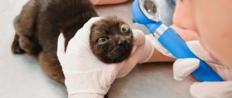
Unfortunately, such a serious pathology does not always have pronounced symptoms, so it can be difficult to recognize even for a veterinarian, not to mention the owner. The disease manifests itself only in the acute stage, while other forms of progression are not noticeable. Therefore, in order to understand that the doctor is dealing with a corneal ulcer, he uses special ophthalmological equipment.
A corneal ulcer may not show any symptoms for some time.
The following symptoms may lead the owner to believe that the cat is developing an eye disease:
- increased tearfulness, due to which the hair around the eyes sticks together, and the eyes themselves seem wet and constantly shiny;
- the eyes become cloudy and take on a red, inflamed tint (which is why the ulcer is often mistaken for conjunctivitis);
- the cat feels uncomfortable - it tries to squint its eyes unnaturally, rub them with its paws (which can only aggravate the course of the disease if the animal transfers additional bacteria to the eye).
A corneal ulcer can be distinguished from conjunctivitis by the method of spread of the disease. If with conjunctivitis in 99 cases out of 100 the disease manifests itself in both eyes, then the ulcer most often affects only one.
Corneal ulceration often causes cats to compulsively scratch the eye.
Pet's well-being
Pets themselves experience corneal ulceration very painfully. However, their behavior will be determined by their individual character. Those cats that openly declare their health begin to meow loudly and protractedly, signaling that their health is not okay. More secretive animals will diligently clean their eyes with their paws, constantly squinting them.
Why is a corneal ulcer dangerous?
Let's say the owner misses the first signs of corneal ulceration, believing that the cat will heal on its own. What will be the results of such an attitude? The longer an animal's cornea ulcerates, the more vessels begin to grow into this same cornea. As a result of this process, sooner or later a hole appears in the eye through which its contents flow out. Accordingly, an untreated corneal ulcer has a good chance of leading to the loss of your pet’s eye.
If the ulcers spread too deep, the animal's eyeball is removed and the eyelids are sutured.
Despite the fact that removing an eyeball sounds like a real death sentence for owners, cats successfully cope with such a difficult operation if they are properly cared for. You can read below about what types of operations exist to remove a cat’s eye and how to care for it in the first days after the incident.
Caring for a cat after eye removal
Keratitis: prevention
Prevention of this disease is quite simple and requires compliance with the following rules:
- Carrying out work in which the eyes may be injured, wearing special safety glasses;
- Strict adherence to hygiene rules when using contact lenses;
- Work with welding in a protective mask, avoiding prolonged exposure to the sun;
- Timely treatment of eye diseases such as: conjunctivitis, blepharitis, dacryocystitis;
- Timely treatment of common pathologies that stimulate the development of keratitis.
If you still notice the first signs of developing this disease, immediately seek professional medical help! Remember: the key to successful treatment is its timeliness!
Diagnostic methods
As mentioned earlier, it is completely impossible to recognize corneal ulceration with the naked eye, so veterinarians resort to special techniques to obtain an unambiguous result.
Superficial ulcers
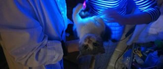
In most cases, doctors prefer to instill a special fluorescent solution into the eyes of cats, which colors the eyeball. After staining, an ultraviolet lamp is placed on the animal’s eye, which allows the injected liquid to be illuminated and any defects on the cornea to be recognized. This method is effective when working with large ulcers, and if the presence of small ulcers is suspected, the veterinarian additionally uses magnifying devices and special filters.
Eye dripped with fluorescent solution
Deep ulcers
If we are talking about deep ulcers, whose course is chronic, then specialists first take material for microscopic examination in order to analyze the characteristics of the ulcer and select a suitable treatment regimen.
Our services in ophthalmology:
The administration of CELT JSC regularly updates the price list posted on the clinic’s website. However, in order to avoid possible misunderstandings, we ask you to clarify the cost of services by phone: +7
| Service name | Price in rubles |
| Appointment with an ophthalmologist (primary) | 3 900 |
| Appointment with a surgical doctor (repeated, for complex programs) within 3 months after the initial one | 2 000 |
| Ultrasound scanning of the anterior segment of the eye | 1 000 |
| Removal of foreign bodies of the cornea, conjunctiva | 2 000 |
All services
Make an appointment through the application or by calling +7 +7 We work every day:
- Monday—Friday: 8.00—20.00
- Saturday: 8.00–18.00
- Sunday is a day off
The nearest metro and MCC stations to the clinic:
- Highway of Enthusiasts or Perovo
- Partisan
- Enthusiast Highway
Driving directions
Treatment
Treatment of corneal ulcers largely depends on the severity of the disease and the depth to which the ulcers have reached. If domestic cats are expected to be seen by a veterinarian at the initial stage of ulceration, then conservative treatment is prescribed. If the animal comes to the doctor in a neglected state, then surgery may be prescribed.
A veterinarian can determine an effective treatment method only after a detailed examination of the animal’s eyes.
The most superficial and small ulcers disappear within a week. If the ulcer reaches Descemet's membrane and a descemetocele occurs, then even the most competent treatment risks not yielding positive results. The owner of the animal must immediately keep in mind that the deeper the ulcers spread, the more expensive it will cost him to treat the animal.
Conservative treatment
In general, treatment for a cat with a corneal ulcer involves the following procedures:
- instilling antibacterial eye drops and/or applying antibacterial ointments to prevent the proliferation of pathogenic bacteria and infections;
- the pet’s use of sedatives or painkillers (they allow the animal to get rid of constant pain, which persists at the time of treatment);
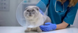
- putting on a protective collar. This protective measure is absolutely necessary, because if the cat periodically scratches its eyes, all treatment will be completely pointless.
Ointments are applied to the cat's eyes using cotton pads or gauze.
Antibiotics
Any antibacterial drops that are prescribed by a veterinarian retain their effect for four hours, after which they stop working. In order for such drops to really help the animal fight the disease, they should be renewed every 3-4 hours.
If you do not have the opportunity to carry out this procedure so often, we recommend turning to ointments. The advantage of antibacterial ointments is that they do not wash out of the eye as quickly as drops, since they are applied to the skin near the eyes.
It is necessary to bury the drops very carefully so as not to cause additional injury to the cat.
Anti-anxiety medications
As a drug that will help a cat cope with pain, veterinarians prefer to prescribe atropine, available in the form of drops. It perfectly relieves pain and spasm, as a result of which the pet stops constantly squinting its eyes. However, this drug also has its own negative side - it helps to dilate the pupil, as a result of which the animal begins to react very painfully to bright light sources.
One percent atropine eye drops
Due to this feature, it is recommended to keep a cat who is prescribed atropine in a room with dim lighting so as not to cause anxiety attacks in him. Moreover, the owner needs to be prepared for the fact that the pupil will regain its previous ability to adjust to lighting only a few days after stopping taking atropine.
Important! Atropine is administered no more than twice a day.
Side effects
Before you start putting drops into your cat’s eyes, you should prepare in advance for the fact that the pet will most likely resist. The point is not only that instillation itself is not easy for animals, but also that antibacterial drops have a bitter, unpleasant taste. Considering that in cats the tear duct has contact with the oral cavity, your pet will have to “feast” on this bitterness for some time.
During eye drops, the cat may show aggression and break out
As a result of taking such medications, the animal may experience increased salivation for the first few minutes due to an unpleasant taste - there is nothing to worry about. Another thing is that with each subsequent instillation, your pet will resist more and more, remembering the past discomfort, and you will have to try hard to instill the solution.
By the way! You can read about how to properly apply eye drops to a cat in a separate article on our portal.
Operation
The operation is prescribed when there is a high probability of losing the eye and drastic measures must be taken as soon as possible. Surgical intervention takes place only for deep ulcers, since superficial variations can be resolved with the help of drops. During the operation, the surgeon performs several actions:
- dissects the damaged cornea;
- applies a flap of unaffected conjunctiva;
- If necessary, sew up the eyelids for a couple of days.
At the end of the operation, a protective collar is immediately put on the cat.
The operation itself takes place under general anesthesia.
It also happens that the ulceration process is accompanied by the accumulation of many dead cells, which interfere with the healing of the eye and the recovery of the pet. In such cases, the surgeon also performs additional removal of accumulated layers of tissue.
To determine the success of the measures taken in the fight against ulceration, the veterinarian prescribes a repeat test with instillation of fluorescent liquid into the animal’s eyes.
Video - Performing surgery on a cat's cornea
Forecast
Timely treatment allows you to preserve vision and prevent complications. During treatment, fluorescein tests are carried out, the results of which determine the degree of healing. Superficial forms epithelialize within 4-5 days. The healing time for deep defects varies from person to person (usually 3-4 weeks). The main factor influencing the recovery period is the treatment of the underlying disease that caused ulcerative keratitis. When it is eliminated, regenerative processes are accelerated.
In our clinic, a doctor is an ophthalmologist, a doctor of the highest category, A. E. Kopenkin .
Videos and Illustrations
Clinical case: Sphynx kitten, corneal ulcer


